👁️ FCPS NCPS IMM MD Paediatrics TOACS
Observed Station | CPSP Format | 8 minutes
⏱️ TIME REMAINING
08:00
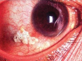
A 2‑year‑old child with failure to thrive, mother concerned for vision.
❓ Q1. Identify the ocular finding shown in the image. What condition is it pathognomonic for?
✅ Model Answer: • Ocular finding: Bitot spot – a foamy, white/yellowish plaque on the conjunctiva.
• Pathognomonic for: Vitamin A deficiency (xerophthalmia).
• Location: Usually on the temporal side of the conjunctiva, often bilateral.
• Appearance: Foamy, white/yellowish, keratinized plaque.
• Pathognomonic for: Vitamin A deficiency (xerophthalmia).
• Location: Usually on the temporal side of the conjunctiva, often bilateral.
• Appearance: Foamy, white/yellowish, keratinized plaque.
❓ Q2. What is the pathophysiology of Bitot spot formation?
✅ Model Answer: • Pathophysiology: Vitamin A deficiency → impaired epithelial differentiation and keratinization.
• Conjunctival epithelium undergoes squamous metaplasia and keratinization.
• Accumulation of desquamated keratinized cells, bacteria, and mucus on the conjunctival surface.
• This forms a foamy, plaque-like lesion – the Bitot spot.
• Bitot spots represent a stage of xerophthalmia (early non-corneal involvement).
• Conjunctival epithelium undergoes squamous metaplasia and keratinization.
• Accumulation of desquamated keratinized cells, bacteria, and mucus on the conjunctival surface.
• This forms a foamy, plaque-like lesion – the Bitot spot.
• Bitot spots represent a stage of xerophthalmia (early non-corneal involvement).
❓ Q3. What are the clinical features and symptoms of vitamin A deficiency?
✅ Model Answer: • Clinical features:
- Night blindness (nyctalopia): Earliest symptom – difficulty seeing in dim light.
- Xerophthalmia: Dryness of the conjunctiva and cornea.
- Bitot spots: Foamy, white/yellowish conjunctival plaques.
- Corneal xerosis: Dry, hazy appearance of the cornea.
- Keratomalacia: Softening and ulceration of the cornea → blindness.
- Systemic: Dry skin (follicular hyperkeratosis), growth failure, impaired immunity, increased susceptibility to infections (especially measles and diarrhea).
- Night blindness (nyctalopia): Earliest symptom – difficulty seeing in dim light.
- Xerophthalmia: Dryness of the conjunctiva and cornea.
- Bitot spots: Foamy, white/yellowish conjunctival plaques.
- Corneal xerosis: Dry, hazy appearance of the cornea.
- Keratomalacia: Softening and ulceration of the cornea → blindness.
- Systemic: Dry skin (follicular hyperkeratosis), growth failure, impaired immunity, increased susceptibility to infections (especially measles and diarrhea).
❓ Q4. What are the risk factors for vitamin A deficiency?
✅ Model Answer: • Risk factors:
- Malnutrition/undernutrition – inadequate dietary intake (poor intake of vitamin A-rich foods).
- Measles – depletes vitamin A stores and increases requirement.
- Diarrhea – leads to malabsorption and increased losses.
- Fat malabsorption – cystic fibrosis, cholestasis, celiac disease.
- Poverty – limited access to vitamin A-rich foods (liver, eggs, dairy, orange vegetables).
- Prematurity – low vitamin A stores at birth.
- Parasitic infections – giardiasis, ascariasis.
- Malnutrition/undernutrition – inadequate dietary intake (poor intake of vitamin A-rich foods).
- Measles – depletes vitamin A stores and increases requirement.
- Diarrhea – leads to malabsorption and increased losses.
- Fat malabsorption – cystic fibrosis, cholestasis, celiac disease.
- Poverty – limited access to vitamin A-rich foods (liver, eggs, dairy, orange vegetables).
- Prematurity – low vitamin A stores at birth.
- Parasitic infections – giardiasis, ascariasis.
❓ Q5. What is the WHO classification of xerophthalmia?
✅ Model Answer: • WHO xerophthalmia classification:
- XN: Night blindness.
- X1A: Conjunctival xerosis (dry conjunctiva).
- X1B: Bitot spots.
- X2: Corneal xerosis (dry, hazy cornea).
- X3A: Corneal ulceration involving less than 1/3 of the cornea.
- X3B: Corneal ulceration involving 1/3 or more of the cornea (keratomalacia).
- XS: Corneal scar (healed keratomalacia).
- XF: Xerophthalmic fundus (rare).
- XN: Night blindness.
- X1A: Conjunctival xerosis (dry conjunctiva).
- X1B: Bitot spots.
- X2: Corneal xerosis (dry, hazy cornea).
- X3A: Corneal ulceration involving less than 1/3 of the cornea.
- X3B: Corneal ulceration involving 1/3 or more of the cornea (keratomalacia).
- XS: Corneal scar (healed keratomalacia).
- XF: Xerophthalmic fundus (rare).
❓ Q6. What is the treatment for a child with Bitot spots due to vitamin A deficiency?
✅ Model Answer: • High-dose vitamin A:
- Dose:
- 6-12 months: 100,000 IU (100,000 IU) orally.
- >12 months: 200,000 IU orally.
- Schedule: Give immediately on diagnosis, repeat the next day, and give a third dose at least 2 weeks later.
- For corneal involvement: Give additional dose on day 14.
• Dietary advice: Encourage vitamin A-rich foods (liver, eggs, dairy, orange/yellow vegetables, dark green leafy vegetables).
• Address underlying malnutrition: Nutritional rehabilitation.
• Treat intercurrent infections: Measles, diarrhea.
- Dose:
- 6-12 months: 100,000 IU (100,000 IU) orally.
- >12 months: 200,000 IU orally.
- Schedule: Give immediately on diagnosis, repeat the next day, and give a third dose at least 2 weeks later.
- For corneal involvement: Give additional dose on day 14.
• Dietary advice: Encourage vitamin A-rich foods (liver, eggs, dairy, orange/yellow vegetables, dark green leafy vegetables).
• Address underlying malnutrition: Nutritional rehabilitation.
• Treat intercurrent infections: Measles, diarrhea.
❓ Q7. Why is vitamin A deficiency associated with increased mortality in measles and diarrhea?
✅ Model Answer: • Vitamin A is essential for:
- Immune function: Maintains epithelial barriers (skin, respiratory, gastrointestinal, genitourinary).
- Mucosal immunity: Deficiency leads to impaired T-cell function, reduced IgA production, and impaired epithelial repair.
- Measles: Measles virus depletes vitamin A stores and damages epithelial surfaces, increasing the risk of severe complications (pneumonia, diarrhea, blindness).
- Diarrhea: Impaired intestinal mucosal integrity leads to more severe and prolonged diarrhea.
- Mortality: Vitamin A deficiency increases the risk of death from measles by 50-75% and from diarrhea by 30-50%.
- Immune function: Maintains epithelial barriers (skin, respiratory, gastrointestinal, genitourinary).
- Mucosal immunity: Deficiency leads to impaired T-cell function, reduced IgA production, and impaired epithelial repair.
- Measles: Measles virus depletes vitamin A stores and damages epithelial surfaces, increasing the risk of severe complications (pneumonia, diarrhea, blindness).
- Diarrhea: Impaired intestinal mucosal integrity leads to more severe and prolonged diarrhea.
- Mortality: Vitamin A deficiency increases the risk of death from measles by 50-75% and from diarrhea by 30-50%.
❓ Q8. What are the dietary sources of vitamin A?
✅ Model Answer: • Preformed vitamin A (retinol):
- Animal sources: Liver (beef, chicken, fish), eggs, dairy products (milk, cheese, butter), fish oils.
• Provitamin A carotenoids (β-carotene):
- Orange/yellow vegetables: Carrots, sweet potatoes, pumpkin, squash.
- Dark green leafy vegetables: Spinach, kale, collard greens, amaranth.
- Orange/yellow fruits: Mangoes, papayas, apricots, cantaloupe.
- Red palm oil.
• Note: β-carotene is converted to vitamin A in the body, but absorption is less efficient than preformed vitamin A.
- Animal sources: Liver (beef, chicken, fish), eggs, dairy products (milk, cheese, butter), fish oils.
• Provitamin A carotenoids (β-carotene):
- Orange/yellow vegetables: Carrots, sweet potatoes, pumpkin, squash.
- Dark green leafy vegetables: Spinach, kale, collard greens, amaranth.
- Orange/yellow fruits: Mangoes, papayas, apricots, cantaloupe.
- Red palm oil.
• Note: β-carotene is converted to vitamin A in the body, but absorption is less efficient than preformed vitamin A.
❓ Q9. What is the role of vitamin A supplementation in public health?
✅ Model Answer: • WHO recommendation: Vitamin A supplementation for children 6-59 months in areas at risk of deficiency.
• Dose: 100,000 IU (6-12 months) or 200,000 IU (12-59 months) every 4-6 months.
• Benefits:
- Reduces all-cause mortality by 12-24%.
- Reduces diarrhea-related mortality by 30-50%.
- Reduces measles-related mortality by 50-75%.
- Prevents xerophthalmia and blindness.
• Integration: Often given with immunization campaigns (e.g., polio, measles) and biannual health days.
• Dose: 100,000 IU (6-12 months) or 200,000 IU (12-59 months) every 4-6 months.
• Benefits:
- Reduces all-cause mortality by 12-24%.
- Reduces diarrhea-related mortality by 30-50%.
- Reduces measles-related mortality by 50-75%.
- Prevents xerophthalmia and blindness.
• Integration: Often given with immunization campaigns (e.g., polio, measles) and biannual health days.
❓ Q10. What is the relationship between measles and vitamin A deficiency?
✅ Model Answer: • Measles and vitamin A deficiency have a bidirectional relationship:
- Measles infection depletes vitamin A stores and increases requirements.
- Vitamin A deficiency impairs the immune response to measles, leading to more severe disease.
- Children with measles and vitamin A deficiency are at higher risk of complications (pneumonia, diarrhea, blindness) and death.
• WHO recommendation: All children with measles should receive two doses of vitamin A (200,000 IU if >12 months, 100,000 IU if 6-12 months) given 24 hours apart, regardless of their vitamin A status.
- Measles infection depletes vitamin A stores and increases requirements.
- Vitamin A deficiency impairs the immune response to measles, leading to more severe disease.
- Children with measles and vitamin A deficiency are at higher risk of complications (pneumonia, diarrhea, blindness) and death.
• WHO recommendation: All children with measles should receive two doses of vitamin A (200,000 IU if >12 months, 100,000 IU if 6-12 months) given 24 hours apart, regardless of their vitamin A status.
❓ Q11. What is the differential diagnosis of Bitot spots?
✅ Model Answer: • Differential diagnoses:
- Pinguecula: Yellowish, raised lesion on the conjunctiva (usually nasal side), not foamy; due to UV exposure and aging.
- Pterygium: Triangular, fleshy growth from the nasal conjunctiva onto the cornea; can be vascularized.
- Conjunctival concretions: Small, hard yellow-white deposits; not foamy.
- Conjunctival papilloma: Raised, fleshy lesion; usually on the bulbar conjunctiva.
- Lipid deposition: Yellowish areas in the conjunctiva due to lipid disorders.
- Key differentiator: Bitot spots are foamy, bilateral, and associated with night blindness and other signs of vitamin A deficiency.
- Pinguecula: Yellowish, raised lesion on the conjunctiva (usually nasal side), not foamy; due to UV exposure and aging.
- Pterygium: Triangular, fleshy growth from the nasal conjunctiva onto the cornea; can be vascularized.
- Conjunctival concretions: Small, hard yellow-white deposits; not foamy.
- Conjunctival papilloma: Raised, fleshy lesion; usually on the bulbar conjunctiva.
- Lipid deposition: Yellowish areas in the conjunctiva due to lipid disorders.
- Key differentiator: Bitot spots are foamy, bilateral, and associated with night blindness and other signs of vitamin A deficiency.
❓ Q12. What is the role of zinc in vitamin A metabolism?
✅ Model Answer: • Zinc and vitamin A interaction:
- Zinc is required for the synthesis of retinol-binding protein (RBP), which transports vitamin A in the blood.
- Zinc deficiency can impair the mobilization and utilization of vitamin A, even if dietary intake is adequate.
- Zinc deficiency can also affect the absorption of vitamin A from the gut.
- Clinical implication: In vitamin A deficiency, zinc supplementation may improve response to vitamin A therapy.
- WHO recommends zinc supplementation for diarrhea in children (10-20 mg/day for 10-14 days), which may also help improve vitamin A status.
- Zinc is required for the synthesis of retinol-binding protein (RBP), which transports vitamin A in the blood.
- Zinc deficiency can impair the mobilization and utilization of vitamin A, even if dietary intake is adequate.
- Zinc deficiency can also affect the absorption of vitamin A from the gut.
- Clinical implication: In vitamin A deficiency, zinc supplementation may improve response to vitamin A therapy.
- WHO recommends zinc supplementation for diarrhea in children (10-20 mg/day for 10-14 days), which may also help improve vitamin A status.
❓ Q13. How would you counsel the parents of a child with Bitot spots?
✅ Model Answer: • "Your child has Bitot spots, which are a sign of vitamin A deficiency. This happens when the diet does not have enough vitamin A."
• "Vitamin A is very important for eye health, growth, and fighting infections. Without enough, children can develop night blindness and, in severe cases, blindness."
• "We will give your child a high dose of vitamin A now, again tomorrow, and again in 2 weeks. This will help correct the deficiency."
• "To prevent this from happening again, we need to improve your child's diet. Foods like eggs, liver, milk, orange vegetables (carrots, sweet potatoes), and green leafy vegetables are good sources."
• "In areas where vitamin A deficiency is common, all children should receive vitamin A supplements every 6 months."
• "Vitamin A is very important for eye health, growth, and fighting infections. Without enough, children can develop night blindness and, in severe cases, blindness."
• "We will give your child a high dose of vitamin A now, again tomorrow, and again in 2 weeks. This will help correct the deficiency."
• "To prevent this from happening again, we need to improve your child's diet. Foods like eggs, liver, milk, orange vegetables (carrots, sweet potatoes), and green leafy vegetables are good sources."
• "In areas where vitamin A deficiency is common, all children should receive vitamin A supplements every 6 months."
❓ Q14. What are the complications of untreated vitamin A deficiency?
✅ Model Answer: • Complications of vitamin A deficiency:
- Ocular: Progression to corneal xerosis, keratomalacia, and irreversible blindness.
- Immunity: Increased susceptibility to infections (measles, diarrhea, respiratory infections).
- Growth: Impaired growth and development.
- Skin: Follicular hyperkeratosis (dry, scaly skin).
- Mortality: Increased risk of death from measles and diarrhea (especially in children under 5 years).
- Anemia: Vitamin A deficiency contributes to iron deficiency anemia by impairing iron mobilization.
- Ocular: Progression to corneal xerosis, keratomalacia, and irreversible blindness.
- Immunity: Increased susceptibility to infections (measles, diarrhea, respiratory infections).
- Growth: Impaired growth and development.
- Skin: Follicular hyperkeratosis (dry, scaly skin).
- Mortality: Increased risk of death from measles and diarrhea (especially in children under 5 years).
- Anemia: Vitamin A deficiency contributes to iron deficiency anemia by impairing iron mobilization.
❓ Q15. What is the role of vitamin A in the management of severe malnutrition?
✅ Model Answer: • Vitamin A in severe acute malnutrition (SAM):
- Children with SAM have depleted vitamin A stores.
- WHO recommends vitamin A supplementation as part of the management of SAM.
- Dose:
- 100,000 IU for 6-12 months
- 200,000 IU for >12 months
- Given on day 1 of treatment, then repeated on day 2 and day 14 (if signs of xerophthalmia).
- Benefits:
- Reduces mortality in severely malnourished children.
- Supports immune function and mucosal integrity.
- Helps prevent eye complications.
- Part of the standard WHO protocol for SAM (along with F-75, F-100, antibiotics, and other micronutrients).
- Children with SAM have depleted vitamin A stores.
- WHO recommends vitamin A supplementation as part of the management of SAM.
- Dose:
- 100,000 IU for 6-12 months
- 200,000 IU for >12 months
- Given on day 1 of treatment, then repeated on day 2 and day 14 (if signs of xerophthalmia).
- Benefits:
- Reduces mortality in severely malnourished children.
- Supports immune function and mucosal integrity.
- Helps prevent eye complications.
- Part of the standard WHO protocol for SAM (along with F-75, F-100, antibiotics, and other micronutrients).
📋 Mock Test Feedback
💡 Examiner's note: Compare your answers with model answers. In real TOACS, you would discuss these with the examiner.