FCPS MCPS IMM MD Paediatrics TOACS
Observed Station | CPSP Format | 8 minutes
⏱️ TIME REMAINING
08:00
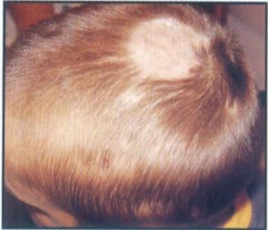
A 5‑year‑old boy is brought to the clinic with a patch of hair loss on the scalp that has been slowly enlarging over the past 2 weeks.
❓ Q1. Identify the scalp condition shown in the image. Describe its characteristic features.
✅ Model Answer: • Condition: Tinea capitis (scalp ringworm) – a dermatophyte infection of the scalp.
• Characteristic features: Patchy alopecia (hair loss), broken hairs at the scalp surface ("black dots"), scaling, erythema, and pruritus. May present with an inflammatory boggy mass (kerion) in some cases. Most common in children aged 3-7 years.
• Characteristic features: Patchy alopecia (hair loss), broken hairs at the scalp surface ("black dots"), scaling, erythema, and pruritus. May present with an inflammatory boggy mass (kerion) in some cases. Most common in children aged 3-7 years.
❓ Q2. What are the most common causative organisms of tinea capitis?
✅ Model Answer: • Most common organisms:
- Trichophyton tonsurans – most common in the US (endothrix, does not fluoresce under Wood lamp).
- Microsporum canis – acquired from pets (dogs, cats); ectothrix, shows green fluorescence under Wood lamp.
- Microsporum audouinii – less common.
- Trichophyton violaceum – endemic in certain regions.
- Microsporum gypseum – soil-associated.
- Trichophyton tonsurans – most common in the US (endothrix, does not fluoresce under Wood lamp).
- Microsporum canis – acquired from pets (dogs, cats); ectothrix, shows green fluorescence under Wood lamp.
- Microsporum audouinii – less common.
- Trichophyton violaceum – endemic in certain regions.
- Microsporum gypseum – soil-associated.
❓ Q3. How is tinea capitis transmitted?
✅ Model Answer: • Transmission:
- Direct contact: With infected humans (T. tonsurans) or animals (M. canis – dogs, cats, rabbits).
- Fomites: Contaminated combs, brushes, hats, hair accessories, barber tools, pillowcases, and towels.
- Asymptomatic carriers: Adults can be asymptomatic carriers and transmit the infection to children.
- Environmental: Soil (Microsporum gypseum).
- Direct contact: With infected humans (T. tonsurans) or animals (M. canis – dogs, cats, rabbits).
- Fomites: Contaminated combs, brushes, hats, hair accessories, barber tools, pillowcases, and towels.
- Asymptomatic carriers: Adults can be asymptomatic carriers and transmit the infection to children.
- Environmental: Soil (Microsporum gypseum).
❓ Q4. What are the clinical variants of tinea capitis?
✅ Model Answer: • Clinical variants:
1. Non-inflammatory (seborrheic-like): Patchy scaling with minimal hair loss, often mistaken for dandruff or seborrheic dermatitis.
2. Black dot tinea capitis: Hairs break off at the scalp surface, leaving "black dots" (T. tonsurans).
3. Gray patch tinea capitis: Well-demarcated, scaly, hypopigmented patches with broken hairs (M. canis).
4. Kerion (inflammatory): Boggy, tender, inflamed mass with pustules and purulent drainage; can lead to scarring alopecia.
5. Favus (tinea favosa): Crusted, cup-shaped yellow crusts (scutula) with a mousy odor; caused by T. schoenleinii (rare).
1. Non-inflammatory (seborrheic-like): Patchy scaling with minimal hair loss, often mistaken for dandruff or seborrheic dermatitis.
2. Black dot tinea capitis: Hairs break off at the scalp surface, leaving "black dots" (T. tonsurans).
3. Gray patch tinea capitis: Well-demarcated, scaly, hypopigmented patches with broken hairs (M. canis).
4. Kerion (inflammatory): Boggy, tender, inflamed mass with pustules and purulent drainage; can lead to scarring alopecia.
5. Favus (tinea favosa): Crusted, cup-shaped yellow crusts (scutula) with a mousy odor; caused by T. schoenleinii (rare).
❓ Q5. What is the role of Wood lamp examination in tinea capitis?
✅ Model Answer: • Wood lamp (Wood's light): A UV light (365 nm) used to detect fungal fluorescence.
• Fluorescence:
- Green fluorescence: Microsporum canis, M. audouinii, M. ferrugineum (ectothrix infections).
- No fluorescence: Trichophyton tonsurans (endothrix – the most common organism in the US).
• Limitations: Not all cases fluoresce; a negative Wood lamp does not rule out tinea capitis. Useful for screening and identifying Microsporum species.
• Fluorescence:
- Green fluorescence: Microsporum canis, M. audouinii, M. ferrugineum (ectothrix infections).
- No fluorescence: Trichophyton tonsurans (endothrix – the most common organism in the US).
• Limitations: Not all cases fluoresce; a negative Wood lamp does not rule out tinea capitis. Useful for screening and identifying Microsporum species.
❓ Q6. What is the diagnostic workup for tinea capitis?
✅ Model Answer: • Diagnostic workup:
1. Clinical examination: Patchy alopecia, broken hairs, scaling, erythema, lymphadenopathy.
2. KOH (potassium hydroxide) preparation: Scrape scales and pluck broken hairs; examine under microscope for arthrospores (within or outside the hair shaft) and hyphae.
3. Wood lamp examination: Green fluorescence suggests Microsporum species.
4. Fungal culture: Gold standard – identifies the organism and guides treatment. Takes 2-3 weeks for growth (sabouraud dextrose agar).
5. Skin biopsy: Rarely needed; may show fungal elements with PAS or GMS staining.
6. PCR: Rapid detection of fungal DNA (available in some centers).
1. Clinical examination: Patchy alopecia, broken hairs, scaling, erythema, lymphadenopathy.
2. KOH (potassium hydroxide) preparation: Scrape scales and pluck broken hairs; examine under microscope for arthrospores (within or outside the hair shaft) and hyphae.
3. Wood lamp examination: Green fluorescence suggests Microsporum species.
4. Fungal culture: Gold standard – identifies the organism and guides treatment. Takes 2-3 weeks for growth (sabouraud dextrose agar).
5. Skin biopsy: Rarely needed; may show fungal elements with PAS or GMS staining.
6. PCR: Rapid detection of fungal DNA (available in some centers).
❓ Q7. What is the treatment of tinea capitis?
✅ Model Answer: • Treatment requires systemic oral antifungal therapy – topical agents are ineffective.
- First-line (Trichophyton tonsurans): Terbinafine (weight-based): 125 mg (<25 kg), 187.5 mg (25-35 kg), 250 mg (>35 kg) daily for 6 weeks.
- Alternative (Microsporum canis): Griseofulvin microsize 20-25 mg/kg/day (max 1 g) for 8-12 weeks (more effective against Microsporum).
- Other options: Itraconazole 5 mg/kg/day for 4-6 weeks; Fluconazole 6 mg/kg/day for 4-6 weeks.
- Adjunctive therapy: Selenium sulfide 2% or ketoconazole 2% shampoo twice weekly to reduce fungal shedding and transmission.
- Kerion: Systemic antifungal + short course prednisone (1-2 mg/kg/day) for 1-2 weeks to reduce inflammation and prevent scarring.
- First-line (Trichophyton tonsurans): Terbinafine (weight-based): 125 mg (<25 kg), 187.5 mg (25-35 kg), 250 mg (>35 kg) daily for 6 weeks.
- Alternative (Microsporum canis): Griseofulvin microsize 20-25 mg/kg/day (max 1 g) for 8-12 weeks (more effective against Microsporum).
- Other options: Itraconazole 5 mg/kg/day for 4-6 weeks; Fluconazole 6 mg/kg/day for 4-6 weeks.
- Adjunctive therapy: Selenium sulfide 2% or ketoconazole 2% shampoo twice weekly to reduce fungal shedding and transmission.
- Kerion: Systemic antifungal + short course prednisone (1-2 mg/kg/day) for 1-2 weeks to reduce inflammation and prevent scarring.
❓ Q8. What is a kerion? How is it managed?
✅ Model Answer: • Kerion: An inflammatory, boggy, tender, swollen mass on the scalp with pustules and purulent drainage. It represents a hypersensitivity reaction to the fungal infection.
• Management:
- Systemic antifungal therapy: Same as for non-inflammatory tinea capitis (terbinafine or griseofulvin).
- Systemic corticosteroids: Prednisone 1-2 mg/kg/day for 1-2 weeks to reduce inflammation, pain, and prevent scarring alopecia.
- Do not incise or drain (may lead to scarring or spread).
- Pain management as needed.
- Culture and sensitivity to guide antifungal choice.
- Follow-up: Close monitoring for resolution and scarring.
• Management:
- Systemic antifungal therapy: Same as for non-inflammatory tinea capitis (terbinafine or griseofulvin).
- Systemic corticosteroids: Prednisone 1-2 mg/kg/day for 1-2 weeks to reduce inflammation, pain, and prevent scarring alopecia.
- Do not incise or drain (may lead to scarring or spread).
- Pain management as needed.
- Culture and sensitivity to guide antifungal choice.
- Follow-up: Close monitoring for resolution and scarring.
❓ Q9. What is the role of selenium sulfide shampoo in tinea capitis?
✅ Model Answer: • Selenium sulfide 2% or ketoconazole 2% shampoo:
- Role: Adjunctive therapy – not curative alone.
- Mechanism: Reduces fungal shedding and spores on the scalp surface.
- Indications: Helps decrease transmission to family members and reduce environmental contamination.
- Use: Twice weekly, leave on for 5-10 minutes, then rinse.
- Benefit: Reduces the duration of infectivity.
- Limitation: Does not penetrate the hair follicle; therefore, systemic antifungal therapy is still required.
- Role: Adjunctive therapy – not curative alone.
- Mechanism: Reduces fungal shedding and spores on the scalp surface.
- Indications: Helps decrease transmission to family members and reduce environmental contamination.
- Use: Twice weekly, leave on for 5-10 minutes, then rinse.
- Benefit: Reduces the duration of infectivity.
- Limitation: Does not penetrate the hair follicle; therefore, systemic antifungal therapy is still required.
❓ Q10. What are the complications of tinea capitis?
✅ Model Answer: • Complications:
- Permanent scarring alopecia: Especially if kerion is untreated or treated inadequately (fibrosis and hair follicle destruction).
- Secondary bacterial infection: Impetigo, cellulitis, abscess formation.
- Kerion with scarring: Can lead to permanent hair loss.
- Psychological impact: Social stigma, anxiety, bullying due to visible hair loss.
- Transmission to family members: Asymptomatic carriers may perpetuate the infection.
- Prolonged course: If treatment is delayed or inappropriate (topical alone).
- Permanent scarring alopecia: Especially if kerion is untreated or treated inadequately (fibrosis and hair follicle destruction).
- Secondary bacterial infection: Impetigo, cellulitis, abscess formation.
- Kerion with scarring: Can lead to permanent hair loss.
- Psychological impact: Social stigma, anxiety, bullying due to visible hair loss.
- Transmission to family members: Asymptomatic carriers may perpetuate the infection.
- Prolonged course: If treatment is delayed or inappropriate (topical alone).
❓ Q11. What is the difference between tinea capitis and alopecia areata?
✅ Model Answer: • Tinea capitis:
- Fungal infection (dermatophyte).
- Scaling, broken hairs ("black dots"), erythema, pruritus.
- May have kerion (inflammatory boggy mass).
- Positive KOH/culture.
- Requires systemic antifungal therapy.
- Wood lamp may show fluorescence (Microsporum).
• Alopecia areata:
- Autoimmune (T-cell mediated) – non-scarring.
- Well-demarcated, smooth, completely bald patches (no scaling, no broken hairs).
- "Exclamation point" hairs at the margins.
- No inflammation or crusting.
- Nail pitting may be present.
- No fungal elements on KOH.
- Treatment: Topical/intralesional steroids, minoxidil, or watchful waiting.
- Fungal infection (dermatophyte).
- Scaling, broken hairs ("black dots"), erythema, pruritus.
- May have kerion (inflammatory boggy mass).
- Positive KOH/culture.
- Requires systemic antifungal therapy.
- Wood lamp may show fluorescence (Microsporum).
• Alopecia areata:
- Autoimmune (T-cell mediated) – non-scarring.
- Well-demarcated, smooth, completely bald patches (no scaling, no broken hairs).
- "Exclamation point" hairs at the margins.
- No inflammation or crusting.
- Nail pitting may be present.
- No fungal elements on KOH.
- Treatment: Topical/intralesional steroids, minoxidil, or watchful waiting.
❓ Q12. What is the role of fungal culture in tinea capitis?
✅ Model Answer: • Fungal culture (gold standard):
- Purpose: Identifies the specific organism (species) causing tinea capitis.
- Method: Pluck hairs (without roots), inoculate onto Sabouraud dextrose agar (with or without cycloheximide/chloramphenicol).
- Growth time: 2-3 weeks (sometimes up to 4 weeks).
- Clinical significance: Guides choice of antifungal therapy (terbinafine for Trichophyton, griseofulvin for Microsporum).
- Monitoring: Follow-up cultures may be needed to confirm cure (especially in resistant cases).
- Limitations: Delayed results; may be negative if patient has been on antifungal therapy.
- Purpose: Identifies the specific organism (species) causing tinea capitis.
- Method: Pluck hairs (without roots), inoculate onto Sabouraud dextrose agar (with or without cycloheximide/chloramphenicol).
- Growth time: 2-3 weeks (sometimes up to 4 weeks).
- Clinical significance: Guides choice of antifungal therapy (terbinafine for Trichophyton, griseofulvin for Microsporum).
- Monitoring: Follow-up cultures may be needed to confirm cure (especially in resistant cases).
- Limitations: Delayed results; may be negative if patient has been on antifungal therapy.
❓ Q13. How would you counsel the parents of a child with tinea capitis?
✅ Model Answer: • "Your child has a fungal infection of the scalp called tinea capitis (scalp ringworm). It is very common in children."
• "This requires oral medication (an antifungal) because the infection is deep in the hair follicles – creams or shampoos alone will not cure it."
• "We will prescribe an antifungal medication (terbinafine or griseofulvin) for 6-8 weeks. It is important to complete the full course, even if the hair starts to grow back."
• "You can also use a medicated shampoo (selenium sulfide or ketoconazole) twice a week to reduce the spread of fungus."
• "Wash combs, brushes, hats, and pillowcases. Check other family members for signs of infection."
• "If the area becomes very swollen, tender, or painful (kerion), we may add a short course of steroid medication to reduce inflammation and prevent scarring."
• "The hair will usually grow back, but scarring may occur if the infection is severe or untreated."
• "This requires oral medication (an antifungal) because the infection is deep in the hair follicles – creams or shampoos alone will not cure it."
• "We will prescribe an antifungal medication (terbinafine or griseofulvin) for 6-8 weeks. It is important to complete the full course, even if the hair starts to grow back."
• "You can also use a medicated shampoo (selenium sulfide or ketoconazole) twice a week to reduce the spread of fungus."
• "Wash combs, brushes, hats, and pillowcases. Check other family members for signs of infection."
• "If the area becomes very swollen, tender, or painful (kerion), we may add a short course of steroid medication to reduce inflammation and prevent scarring."
• "The hair will usually grow back, but scarring may occur if the infection is severe or untreated."
❓ Q14. What is the difference between endothrix and ectothrix infections?
✅ Model Answer: • Endothrix (Trichophyton tonsurans):
- Arthrospores are within the hair shaft.
- Hair shaft is not surrounded by fungal elements.
- Hair breaks off at the scalp surface → "black dots".
- No Wood lamp fluorescence.
- Common in the US.
• Ectothrix (Microsporum canis, M. audouinii):
- Arthrospores are outside the hair shaft (sheath of spores).
- Hair shaft is surrounded by fungal elements.
- Hair breaks off above the scalp surface → gray patch.
- Green Wood lamp fluorescence.
- Common in animal-associated infections.
- Arthrospores are within the hair shaft.
- Hair shaft is not surrounded by fungal elements.
- Hair breaks off at the scalp surface → "black dots".
- No Wood lamp fluorescence.
- Common in the US.
• Ectothrix (Microsporum canis, M. audouinii):
- Arthrospores are outside the hair shaft (sheath of spores).
- Hair shaft is surrounded by fungal elements.
- Hair breaks off above the scalp surface → gray patch.
- Green Wood lamp fluorescence.
- Common in animal-associated infections.
❓ Q15. What is the prognosis for a child with tinea capitis?
✅ Model Answer: • Prognosis: Excellent with appropriate treatment.
- Cure rates: >90% with proper oral antifungal therapy.
- Hair regrowth: Usually begins within 4-6 weeks of starting treatment; full regrowth may take several months.
- Scarring: Rare if treated early; kerion can lead to permanent scarring alopecia if not treated promptly with corticosteroids.
- Relapse: Uncommon but can occur if treatment is not completed or if reinfection occurs.
- Carriers: Asymptomatic carriers (especially adults) should be identified and treated to prevent reinfection.
- Follow-up: Clinical and fungal culture follow-up may be needed to confirm cure.
- Cure rates: >90% with proper oral antifungal therapy.
- Hair regrowth: Usually begins within 4-6 weeks of starting treatment; full regrowth may take several months.
- Scarring: Rare if treated early; kerion can lead to permanent scarring alopecia if not treated promptly with corticosteroids.
- Relapse: Uncommon but can occur if treatment is not completed or if reinfection occurs.
- Carriers: Asymptomatic carriers (especially adults) should be identified and treated to prevent reinfection.
- Follow-up: Clinical and fungal culture follow-up may be needed to confirm cure.
📋 Mock Test Feedback
💡 Examiner's note: Compare your answers with model answers. In real TOACS, you would discuss these with the examiner.