FCPS MCPS IMM MD Paediatrics TOACS
Observed Station | CPSP Format | 8 minutes
⏱️ TIME REMAINING
08:00
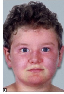
A 6‑year‑old boy is brought in with a reddish-purple rash on his face that has been present for 2 months. His mother reports he has been fatigued and has had muscle pain.
❓ Q1. Identify the skin rash shown in the image. What condition is it pathognomonic for?
✅ Model Answer: • Skin rash: Heliotrope rash – erythematous to violaceous discoloration involving the periorbital area, eyelids, and malar eminences.
• Pathognomonic for: Juvenile dermatomyositis (JDM).
• Key features: Photosensitive, may spare nasolabial folds (unlike SLE), often associated with Gottron papules and proximal muscle weakness.
• Pathognomonic for: Juvenile dermatomyositis (JDM).
• Key features: Photosensitive, may spare nasolabial folds (unlike SLE), often associated with Gottron papules and proximal muscle weakness.
❓ Q2. What is the pathophysiology of juvenile dermatomyositis?
✅ Model Answer: • Pathophysiology: JDM is an immune-mediated vasculopathy affecting the skin, muscles, and gastrointestinal tract.
• Mechanism: Complement-mediated microangiopathy (C5b-9 membrane attack complex) deposits in the endomysial capillaries → capillary damage → muscle ischemia and necrosis.
• Inflammation: CD4+ T cells, macrophages, and B cells infiltrate the perivascular and perimysial areas.
• Myositis-specific antibodies: Associated with specific phenotypes (e.g., TIF1γ, NXP2, MDA5).
• Mechanism: Complement-mediated microangiopathy (C5b-9 membrane attack complex) deposits in the endomysial capillaries → capillary damage → muscle ischemia and necrosis.
• Inflammation: CD4+ T cells, macrophages, and B cells infiltrate the perivascular and perimysial areas.
• Myositis-specific antibodies: Associated with specific phenotypes (e.g., TIF1γ, NXP2, MDA5).
❓ Q3. What are the diagnostic criteria for juvenile dermatomyositis?
✅ Model Answer: • Diagnostic criteria (Bohan & Peter 1975):
1. Heliotrope rash – violaceous erythema on eyelids.
2. Gottron papules – erythematous, scaly papules over knuckles, elbows, knees.
3. Proximal muscle weakness – symmetric weakness of shoulder and pelvic girdle muscles.
4. Elevated muscle enzymes – CK, aldolase, AST, ALT, LDH.
5. EMG changes – myopathic pattern with fibrillation potentials.
6. Muscle biopsy – perifascicular atrophy, capillary depletion.
• Definite JDM: Rash + ≥3 of the remaining criteria.
• Probable JDM: Rash + 2 criteria.
• MRI (STIR showing muscle edema) is now often used instead of EMG/biopsy.
1. Heliotrope rash – violaceous erythema on eyelids.
2. Gottron papules – erythematous, scaly papules over knuckles, elbows, knees.
3. Proximal muscle weakness – symmetric weakness of shoulder and pelvic girdle muscles.
4. Elevated muscle enzymes – CK, aldolase, AST, ALT, LDH.
5. EMG changes – myopathic pattern with fibrillation potentials.
6. Muscle biopsy – perifascicular atrophy, capillary depletion.
• Definite JDM: Rash + ≥3 of the remaining criteria.
• Probable JDM: Rash + 2 criteria.
• MRI (STIR showing muscle edema) is now often used instead of EMG/biopsy.
❓ Q4. What are the cutaneous manifestations of juvenile dermatomyositis?
✅ Model Answer: • Cutaneous manifestations:
- Heliotrope rash: Purple-red discoloration of eyelids and periorbital area; pathognomonic.
- Gottron papules: Erythematous, scaly papules over the extensor surfaces of knuckles, elbows, knees, and medial malleoli.
- Gottron sign: Confluent erythema over the knuckles (without papules).
- Malar rash: Erythematous to violaceous rash on cheeks (similar to SLE but may spare nasolabial folds).
- Photosensitivity.
- Periungual telangiectasias: Dilated capillaries around the nail folds.
- Mechanic's hands: Thickened, scaly, fissured skin on the palmar surface of fingers (associated with anti-MDA5).
- Calcinosis: Subcutaneous calcium deposits (late complication, especially with delayed treatment).
- Heliotrope rash: Purple-red discoloration of eyelids and periorbital area; pathognomonic.
- Gottron papules: Erythematous, scaly papules over the extensor surfaces of knuckles, elbows, knees, and medial malleoli.
- Gottron sign: Confluent erythema over the knuckles (without papules).
- Malar rash: Erythematous to violaceous rash on cheeks (similar to SLE but may spare nasolabial folds).
- Photosensitivity.
- Periungual telangiectasias: Dilated capillaries around the nail folds.
- Mechanic's hands: Thickened, scaly, fissured skin on the palmar surface of fingers (associated with anti-MDA5).
- Calcinosis: Subcutaneous calcium deposits (late complication, especially with delayed treatment).
❓ Q5. What are the muscular manifestations of juvenile dermatomyositis?
✅ Model Answer: • Muscular manifestations:
- Proximal muscle weakness: Symmetric weakness of shoulder girdle (difficulty lifting arms, combing hair) and pelvic girdle (difficulty climbing stairs, rising from the floor).
- Gower sign: Child uses hands to "walk up" the legs when rising from a squat or lying on the floor.
- Myalgia: Muscle pain and tenderness.
- Fatigue: Often profound.
- Dysphagia: Difficulty swallowing (esophageal involvement) – a sign of more severe disease.
- Respiratory muscle weakness: Can lead to restrictive lung disease.
- Proximal muscle weakness: Symmetric weakness of shoulder girdle (difficulty lifting arms, combing hair) and pelvic girdle (difficulty climbing stairs, rising from the floor).
- Gower sign: Child uses hands to "walk up" the legs when rising from a squat or lying on the floor.
- Myalgia: Muscle pain and tenderness.
- Fatigue: Often profound.
- Dysphagia: Difficulty swallowing (esophageal involvement) – a sign of more severe disease.
- Respiratory muscle weakness: Can lead to restrictive lung disease.
❓ Q6. What is the role of muscle enzymes in diagnosing JDM?
✅ Model Answer: • Muscle enzymes:
- Creatine kinase (CK): Most specific and sensitive for muscle damage; elevated in ~80% of JDM patients.
- Aldolase: More sensitive than CK, often elevated even when CK is normal.
- AST, ALT, LDH: Can be elevated due to muscle involvement (not just liver).
- Correlation: Enzyme levels often correlate with disease activity; they decrease with treatment.
- Limitations: Can be normal in early disease or with muscle atrophy; not specific to JDM.
- Creatine kinase (CK): Most specific and sensitive for muscle damage; elevated in ~80% of JDM patients.
- Aldolase: More sensitive than CK, often elevated even when CK is normal.
- AST, ALT, LDH: Can be elevated due to muscle involvement (not just liver).
- Correlation: Enzyme levels often correlate with disease activity; they decrease with treatment.
- Limitations: Can be normal in early disease or with muscle atrophy; not specific to JDM.
❓ Q7. What is the role of MRI in diagnosing JDM?
✅ Model Answer: • MRI role:
- STIR (short tau inversion recovery) sequence: Shows muscle edema (inflammation) – a hallmark of active JDM.
- Location: Often shows bilateral, symmetric involvement of proximal muscles (thighs, shoulders).
- Advantages: Non-invasive, more sensitive than EMG, can guide biopsy site.
- Follow-up: Can monitor response to treatment.
- Specificity: High sensitivity but not specific to JDM (can be seen in other myopathies).
- STIR (short tau inversion recovery) sequence: Shows muscle edema (inflammation) – a hallmark of active JDM.
- Location: Often shows bilateral, symmetric involvement of proximal muscles (thighs, shoulders).
- Advantages: Non-invasive, more sensitive than EMG, can guide biopsy site.
- Follow-up: Can monitor response to treatment.
- Specificity: High sensitivity but not specific to JDM (can be seen in other myopathies).
❓ Q8. What is the first-line treatment for juvenile dermatomyositis?
✅ Model Answer: • First-line treatment:
- Corticosteroids: Prednisone 2 mg/kg/day (max 60-80 mg) or IV methylprednisolone 30 mg/kg/day for 3 days (pulse therapy) in severe disease.
- Steroid-sparing agent: Methotrexate 15-20 mg/m²/week (subcutaneous or oral) – started early to reduce steroid burden and prevent calcinosis.
- Duration: Steroids are tapered slowly over 1-2 years (avoid rapid withdrawal).
- Sun protection: Essential to prevent photosensitivity flares.
- Physical therapy: To maintain muscle strength and prevent contractures.
- Corticosteroids: Prednisone 2 mg/kg/day (max 60-80 mg) or IV methylprednisolone 30 mg/kg/day for 3 days (pulse therapy) in severe disease.
- Steroid-sparing agent: Methotrexate 15-20 mg/m²/week (subcutaneous or oral) – started early to reduce steroid burden and prevent calcinosis.
- Duration: Steroids are tapered slowly over 1-2 years (avoid rapid withdrawal).
- Sun protection: Essential to prevent photosensitivity flares.
- Physical therapy: To maintain muscle strength and prevent contractures.
❓ Q9. What are the myositis-specific antibodies in JDM and their associations?
✅ Model Answer: • Myositis-specific antibodies:
- Anti-TIF1γ: Associated with severe, extensive rash, photosensitivity, lipodystrophy, and higher risk of malignancy (rare in children).
- Anti-NXP2: Associated with calcinosis (subcutaneous calcium deposits), and more severe disease.
- Anti-MDA5: Associated with rapidly progressive interstitial lung disease (ILD), skin ulcers, and mechanic's hands.
- Anti-Mi-2: Associated with classic JDM (rash + myositis), good response to steroids.
- Anti-SRP: Associated with severe myositis, poor response to steroids (more common in adults).
- Anti-Jo-1: Associated with ILD, arthritis (more common in adults with antisynthetase syndrome).
- Anti-TIF1γ: Associated with severe, extensive rash, photosensitivity, lipodystrophy, and higher risk of malignancy (rare in children).
- Anti-NXP2: Associated with calcinosis (subcutaneous calcium deposits), and more severe disease.
- Anti-MDA5: Associated with rapidly progressive interstitial lung disease (ILD), skin ulcers, and mechanic's hands.
- Anti-Mi-2: Associated with classic JDM (rash + myositis), good response to steroids.
- Anti-SRP: Associated with severe myositis, poor response to steroids (more common in adults).
- Anti-Jo-1: Associated with ILD, arthritis (more common in adults with antisynthetase syndrome).
❓ Q10. What are the complications of juvenile dermatomyositis?
✅ Model Answer: • Complications:
- Calcinosis: Subcutaneous calcium deposits (most common late complication, especially with delayed treatment).
- Lipodystrophy: Loss of subcutaneous fat, usually upper body (associated with anti-TIF1γ).
- Gastrointestinal vasculitis: Can lead to bowel perforation, GI bleeding, and peritonitis (emergency).
- Interstitial lung disease (ILD): Especially with anti-MDA5; can be rapidly progressive.
- Joint contractures: Due to muscle weakness and prolonged immobility.
- Dysphagia: Can lead to aspiration pneumonia.
- Cardiac involvement: Myocarditis, arrhythmias (rare).
- Growth retardation: Due to chronic corticosteroid use.
- Calcinosis: Subcutaneous calcium deposits (most common late complication, especially with delayed treatment).
- Lipodystrophy: Loss of subcutaneous fat, usually upper body (associated with anti-TIF1γ).
- Gastrointestinal vasculitis: Can lead to bowel perforation, GI bleeding, and peritonitis (emergency).
- Interstitial lung disease (ILD): Especially with anti-MDA5; can be rapidly progressive.
- Joint contractures: Due to muscle weakness and prolonged immobility.
- Dysphagia: Can lead to aspiration pneumonia.
- Cardiac involvement: Myocarditis, arrhythmias (rare).
- Growth retardation: Due to chronic corticosteroid use.
❓ Q11. How does the malar rash in JDM differ from the malar rash in SLE?
✅ Model Answer: • JDM malar rash:
- Erythematous to violaceous rash, may involve the eyelids (heliotrope).
- Often spares the nasolabial folds (a key differentiator).
- Associated with Gottron papules (scaly papules over knuckles).
- Associated with proximal muscle weakness and elevated muscle enzymes.
- Photosensitive.
• SLE malar rash:
- Erythematous, often spares nasolabial folds (classic butterfly rash).
- No associated Gottron papules or muscle weakness.
- Associated with systemic symptoms (arthritis, serositis, renal involvement, hematologic abnormalities).
- Positive ANA, anti-dsDNA, anti-Smith, etc.
- Erythematous to violaceous rash, may involve the eyelids (heliotrope).
- Often spares the nasolabial folds (a key differentiator).
- Associated with Gottron papules (scaly papules over knuckles).
- Associated with proximal muscle weakness and elevated muscle enzymes.
- Photosensitive.
• SLE malar rash:
- Erythematous, often spares nasolabial folds (classic butterfly rash).
- No associated Gottron papules or muscle weakness.
- Associated with systemic symptoms (arthritis, serositis, renal involvement, hematologic abnormalities).
- Positive ANA, anti-dsDNA, anti-Smith, etc.
❓ Q12. What is the role of IVIG in the treatment of JDM?
✅ Model Answer: • IVIG (intravenous immunoglobulin):
- Indications: Used as a steroid-sparing agent in patients who are resistant to corticosteroids and methotrexate, or those with severe disease (including dysphagia, GI vasculitis, or rapidly progressive disease).
- Mechanism: Modulates immune response, neutralizes pathogenic antibodies, and reduces complement deposition.
- Dose: 2 g/kg per month (usually given over 2-5 days).
- Efficacy: Shown to improve muscle strength and skin rash in refractory JDM.
- Side effects: Headache, aseptic meningitis, thrombosis, and renal impairment.
- Indications: Used as a steroid-sparing agent in patients who are resistant to corticosteroids and methotrexate, or those with severe disease (including dysphagia, GI vasculitis, or rapidly progressive disease).
- Mechanism: Modulates immune response, neutralizes pathogenic antibodies, and reduces complement deposition.
- Dose: 2 g/kg per month (usually given over 2-5 days).
- Efficacy: Shown to improve muscle strength and skin rash in refractory JDM.
- Side effects: Headache, aseptic meningitis, thrombosis, and renal impairment.
❓ Q13. How would you counsel the parents of a child with JDM?
✅ Model Answer: • "Your child has juvenile dermatomyositis, a condition that causes inflammation of the skin and muscles. It is an autoimmune disease, meaning the body's immune system is attacking the muscles and skin."
• "The rash (heliotrope) and muscle weakness are hallmark features. We will treat it with medications to control the inflammation, starting with steroids and methotrexate."
• "Early and aggressive treatment is very important to prevent long-term complications like calcium deposits in the skin (calcinosis)."
• "Your child will need regular follow-up with a rheumatologist, physical therapy to maintain muscle strength, and sun protection to prevent the rash from worsening."
• "With appropriate treatment, most children improve significantly, though treatment may last for 1-2 years."
• "Please monitor for signs of difficulty swallowing, severe abdominal pain, or breathing problems – these are emergencies."
• "The rash (heliotrope) and muscle weakness are hallmark features. We will treat it with medications to control the inflammation, starting with steroids and methotrexate."
• "Early and aggressive treatment is very important to prevent long-term complications like calcium deposits in the skin (calcinosis)."
• "Your child will need regular follow-up with a rheumatologist, physical therapy to maintain muscle strength, and sun protection to prevent the rash from worsening."
• "With appropriate treatment, most children improve significantly, though treatment may last for 1-2 years."
• "Please monitor for signs of difficulty swallowing, severe abdominal pain, or breathing problems – these are emergencies."
❓ Q14. What is the role of methotrexate in JDM?
✅ Model Answer: • Methotrexate (MTX) role:
- Indications: First-line steroid-sparing agent in JDM, started early to reduce corticosteroid burden and prevent calcinosis.
- Mechanism: Antifolate and anti-inflammatory agent; suppresses T-cell proliferation and cytokine production.
- Dose: 15-20 mg/m²/week (subcutaneous or oral).
- Monitoring: Complete blood count, liver function tests, renal function every 4-8 weeks.
- Supplementation: Folic acid 1 mg daily to reduce side effects (GI upset, hepatotoxicity).
- Duration: Often continued for 1-2 years after achieving remission.
- Indications: First-line steroid-sparing agent in JDM, started early to reduce corticosteroid burden and prevent calcinosis.
- Mechanism: Antifolate and anti-inflammatory agent; suppresses T-cell proliferation and cytokine production.
- Dose: 15-20 mg/m²/week (subcutaneous or oral).
- Monitoring: Complete blood count, liver function tests, renal function every 4-8 weeks.
- Supplementation: Folic acid 1 mg daily to reduce side effects (GI upset, hepatotoxicity).
- Duration: Often continued for 1-2 years after achieving remission.
❓ Q15. What is the prognosis for a child with JDM?
✅ Model Answer: • Prognosis: Highly variable; depends on the severity of disease, presence of complications, and response to treatment.
- Overall: With early aggressive treatment, ~80% of children achieve remission.
- Risk factors for poor prognosis: Anti-MDA5 (ILD), anti-NXP2 (calcinosis), delayed treatment, dysphagia, GI vasculitis.
- Calcinosis: Develops in ~30-50% of patients, especially if treatment is delayed.
- Relapses: Can occur, especially during medication withdrawal; long-term follow-up is essential.
- Mortality: Low (<2%) with modern treatment, but can occur due to GI perforation, ILD, or cardiac involvement.
- Quality of life: Good with multidisciplinary care and physical rehabilitation.
- Overall: With early aggressive treatment, ~80% of children achieve remission.
- Risk factors for poor prognosis: Anti-MDA5 (ILD), anti-NXP2 (calcinosis), delayed treatment, dysphagia, GI vasculitis.
- Calcinosis: Develops in ~30-50% of patients, especially if treatment is delayed.
- Relapses: Can occur, especially during medication withdrawal; long-term follow-up is essential.
- Mortality: Low (<2%) with modern treatment, but can occur due to GI perforation, ILD, or cardiac involvement.
- Quality of life: Good with multidisciplinary care and physical rehabilitation.
📋 Mock Test Feedback
💡 Examiner's note: Compare your answers with model answers. In real TOACS, you would discuss these with the examiner.