⚕️ FCPS Paediatrics TOACS · Mock Test
| Observed Station | CPSP Format
⏱️ TIME REMAINING
08:00
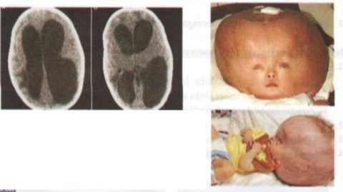
❓ Q1. Describe the findings in the image. What is the most likely diagnosis? List the clinical signs of increased ICP in a newborn.
✅ Model Answer:
• Findings: Macrocephaly (head circumference crossing percentiles), prominent scalp veins, splayed cranial sutures, tense anterior fontanel, setting-sun sign.
• Diagnosis: Congenital hydrocephalus.
• Signs of increased ICP in newborn: Rapid head growth, bulging fontanel, splayed sutures, sunset sign, irritability, vomiting, poor feeding, high-pitched cry, lethargy, apnea.
• Findings: Macrocephaly (head circumference crossing percentiles), prominent scalp veins, splayed cranial sutures, tense anterior fontanel, setting-sun sign.
• Diagnosis: Congenital hydrocephalus.
• Signs of increased ICP in newborn: Rapid head growth, bulging fontanel, splayed sutures, sunset sign, irritability, vomiting, poor feeding, high-pitched cry, lethargy, apnea.
❓ Q2. Define hydrocephalus. What is the difference between communicating and non-communicating (obstructive) hydrocephalus?
✅ Model Answer:
• Hydrocephalus: Abnormal accumulation of CSF within ventricles (ventriculomegaly) due to impaired flow, absorption, or rarely overproduction.
• Non-communicating (obstructive): Obstruction within ventricular system (aqueductal stenosis, foramen of Monro atresia). Most common congenital type.
• Communicating: No obstruction; CSF flows out but absorption impaired (post-hemorrhagic, post-meningitic, Chiari, Dandy-Walker).
• Hydrocephalus: Abnormal accumulation of CSF within ventricles (ventriculomegaly) due to impaired flow, absorption, or rarely overproduction.
• Non-communicating (obstructive): Obstruction within ventricular system (aqueductal stenosis, foramen of Monro atresia). Most common congenital type.
• Communicating: No obstruction; CSF flows out but absorption impaired (post-hemorrhagic, post-meningitic, Chiari, Dandy-Walker).
❓ Q3. What are the common causes of congenital hydrocephalus?
✅ Model Answer:
• Aqueductal stenosis (most common, 40%) – X-linked (L1CAM) or sporadic.
• Chiari II malformation (with myelomeningocele).
• Dandy-Walker malformation.
• Post-hemorrhagic (IVH in preterm infants).
• Post-infectious (congenital CMV, toxoplasmosis, neonatal meningitis).
• Vein of Galen malformation.
• Chromosomal anomalies (trisomy 13,18,21).
• Aqueductal stenosis (most common, 40%) – X-linked (L1CAM) or sporadic.
• Chiari II malformation (with myelomeningocele).
• Dandy-Walker malformation.
• Post-hemorrhagic (IVH in preterm infants).
• Post-infectious (congenital CMV, toxoplasmosis, neonatal meningitis).
• Vein of Galen malformation.
• Chromosomal anomalies (trisomy 13,18,21).
❓ Q4. What imaging is indicated to diagnose hydrocephalus? What are the findings?
✅ Model Answer:
• First-line: Cranial ultrasound (through fontanel) – shows ventriculomegaly, periventricular lucency (edema), IVH, aqueductal stenosis.
• MRI (gold standard) – identifies level of obstruction, associated anomalies (Chiari, Dandy-Walker).
• Findings: Dilation of lateral ventricles ("Mickey Mouse"), transependymal CSF flow, third ventricle dilation.
• First-line: Cranial ultrasound (through fontanel) – shows ventriculomegaly, periventricular lucency (edema), IVH, aqueductal stenosis.
• MRI (gold standard) – identifies level of obstruction, associated anomalies (Chiari, Dandy-Walker).
• Findings: Dilation of lateral ventricles ("Mickey Mouse"), transependymal CSF flow, third ventricle dilation.
❓ Q5. What is the medical (non-surgical) management of hydrocephalus? Is it effective?
✅ Model Answer:
• Medical therapy rarely definitive – temporary or for mild non-progressive hydrocephalus.
• Acetazolamide (carbonic anhydrase inhibitor) – reduces CSF production by 50%. Side effects: metabolic acidosis.
• Furosemide – may be added.
• Serial lumbar punctures – for post-hemorrhagic hydrocephalus in preterm infants.
• Surgery is definitive for progressive hydrocephalus with increased ICP signs.
• Medical therapy rarely definitive – temporary or for mild non-progressive hydrocephalus.
• Acetazolamide (carbonic anhydrase inhibitor) – reduces CSF production by 50%. Side effects: metabolic acidosis.
• Furosemide – may be added.
• Serial lumbar punctures – for post-hemorrhagic hydrocephalus in preterm infants.
• Surgery is definitive for progressive hydrocephalus with increased ICP signs.
❓ Q6. What are the indications for VP shunt insertion in congenital hydrocephalus?
✅ Model Answer:
• Absolute indications: Progressive ventriculomegaly, signs of increased ICP (bulging fontanel, sunset sign, vomiting, irritability, apnea), rapid head growth crossing percentiles.
• Procedure: Ventricular catheter to lateral ventricle → unidirectional valve → peritoneal catheter (VP shunt).
• Goal: Divert CSF to peritoneal cavity for absorption.
• Absolute indications: Progressive ventriculomegaly, signs of increased ICP (bulging fontanel, sunset sign, vomiting, irritability, apnea), rapid head growth crossing percentiles.
• Procedure: Ventricular catheter to lateral ventricle → unidirectional valve → peritoneal catheter (VP shunt).
• Goal: Divert CSF to peritoneal cavity for absorption.
❓ Q7. What is endoscopic third ventriculostomy (ETV)? When is it preferred over a VP shunt?
✅ Model Answer:
• ETV: Endoscopic fenestration in floor of third ventricle → CSF flows directly to subarachnoid space. No shunt hardware.
• Best candidates: Non-communicating hydrocephalus, especially aqueductal stenosis.
• Advantages: No shunt complications (infection, obstruction, over-drainage).
• Success rate: 60-80% for aqueductal stenosis; lower in infants <6 months.
• ETV failure requires VP shunt.
• ETV: Endoscopic fenestration in floor of third ventricle → CSF flows directly to subarachnoid space. No shunt hardware.
• Best candidates: Non-communicating hydrocephalus, especially aqueductal stenosis.
• Advantages: No shunt complications (infection, obstruction, over-drainage).
• Success rate: 60-80% for aqueductal stenosis; lower in infants <6 months.
• ETV failure requires VP shunt.
❓ Q8. What are the common complications of VP shunts? How do they present?
✅ Model Answer:
• Infection (5-10%): Staph epidermidis/aureus. Fever, irritability, erythema/swelling along shunt tract. Requires shunt removal, EVD, IV antibiotics.
• Obstruction (most common, 30-40%): Presents with recurrent increased ICP signs. Diagnosis: shunt series X-ray, CT/MRI, shunt tap.
• Over-drainage (slit ventricle syndrome): Collapsed ventricles, intermittent headaches.
• Abdominal complications: Perforation, pseudocyst, ascites.
• Infection (5-10%): Staph epidermidis/aureus. Fever, irritability, erythema/swelling along shunt tract. Requires shunt removal, EVD, IV antibiotics.
• Obstruction (most common, 30-40%): Presents with recurrent increased ICP signs. Diagnosis: shunt series X-ray, CT/MRI, shunt tap.
• Over-drainage (slit ventricle syndrome): Collapsed ventricles, intermittent headaches.
• Abdominal complications: Perforation, pseudocyst, ascites.
❓ Q9. How would you recognize a VP shunt malfunction in an infant with previously well-controlled hydrocephalus?
✅ Model Answer:
• Symptoms: Irritability, vomiting, lethargy, poor feeding, high-pitched cry, sunset sign, bulging fontanel, rapid head growth.
• Physical signs: Tense fontanel, splayed sutures, prominent scalp veins.
• Imaging: CT/MRI shows increased ventricular size compared to baseline.
• Shunt series X-ray: Look for catheter disconnection or kinking.
• Any infant with VP shunt and new vomiting/irritability = presumed shunt malfunction until proven otherwise.
• Symptoms: Irritability, vomiting, lethargy, poor feeding, high-pitched cry, sunset sign, bulging fontanel, rapid head growth.
• Physical signs: Tense fontanel, splayed sutures, prominent scalp veins.
• Imaging: CT/MRI shows increased ventricular size compared to baseline.
• Shunt series X-ray: Look for catheter disconnection or kinking.
• Any infant with VP shunt and new vomiting/irritability = presumed shunt malfunction until proven otherwise.
❓ Q10. Can congenital hydrocephalus be diagnosed prenatally? What are the ultrasound findings?
✅ Model Answer:
• Yes, diagnosed prenatally by ultrasound (second trimester).
• Findings: Ventriculomegaly (lateral ventricular atrium >10 mm at 18-22 weeks), dangling choroid plexus, thinning of cerebral mantle.
• Associated anomalies: Neural tube defects, Dandy-Walker, aqueductal stenosis, Chiari.
• Management: Fetal MRI, echocardiogram, amniocentesis. Deliver at tertiary center with neurosurgery.
• Yes, diagnosed prenatally by ultrasound (second trimester).
• Findings: Ventriculomegaly (lateral ventricular atrium >10 mm at 18-22 weeks), dangling choroid plexus, thinning of cerebral mantle.
• Associated anomalies: Neural tube defects, Dandy-Walker, aqueductal stenosis, Chiari.
• Management: Fetal MRI, echocardiogram, amniocentesis. Deliver at tertiary center with neurosurgery.
❓ Q11. What is the neurodevelopmental prognosis for an infant with congenital hydrocephalus treated with VP shunt?
✅ Model Answer:
• Depends on etiology, timing, associated anomalies. Isolated aqueductal stenosis → 70-80% normal cognition.
• Sequelae: Cognitive deficits (30-50% learning disability or intellectual disability), spastic diplegia/quadriplegia, strabismus, epilepsy (15-20%), precocious puberty.
• Long-term multidisciplinary follow-up needed (neurosurgery, neurology, developmental pediatrics, ophthalmology).
• Depends on etiology, timing, associated anomalies. Isolated aqueductal stenosis → 70-80% normal cognition.
• Sequelae: Cognitive deficits (30-50% learning disability or intellectual disability), spastic diplegia/quadriplegia, strabismus, epilepsy (15-20%), precocious puberty.
• Long-term multidisciplinary follow-up needed (neurosurgery, neurology, developmental pediatrics, ophthalmology).
❓ Q12. How will you counsel the parents of this infant with congenital hydrocephalus regarding the need for VP shunt and long-term outcome?
✅ Model Answer:
• “Your baby has hydrocephalus – fluid buildup in brain ventricles causing head enlargement and pressure. Not your fault.”
• “Treatment: Shunt surgery to drain fluid from brain to belly. This is done by a pediatric neurosurgeon.”
• “Shunts can get blocked or infected (10% infection risk). Watch for vomiting, irritability, bulging soft spot, sunset eyes – come immediately if these occur.”
• “Outlook varies. Many have normal intelligence. Some may have learning disabilities, motor problems, vision issues, or seizures. Early treatment improves outcomes.”
• “Your baby will need long-term follow-up with multiple specialists.”
• “Your baby has hydrocephalus – fluid buildup in brain ventricles causing head enlargement and pressure. Not your fault.”
• “Treatment: Shunt surgery to drain fluid from brain to belly. This is done by a pediatric neurosurgeon.”
• “Shunts can get blocked or infected (10% infection risk). Watch for vomiting, irritability, bulging soft spot, sunset eyes – come immediately if these occur.”
• “Outlook varies. Many have normal intelligence. Some may have learning disabilities, motor problems, vision issues, or seizures. Early treatment improves outcomes.”
• “Your baby will need long-term follow-up with multiple specialists.”
📋 Mock Test Feedback
💡 Examiner's note: Compare your answers with the model answers. In real TOACS, you would discuss these with the examiner.