⚕️ FCPS Paediatrics TOACS · Mock Test
| Observed Station | CPSP Format
⏱️ TIME REMAINING
08:00
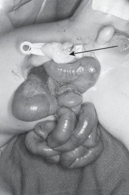
❓ Q1. Describe the findings in the image. What is the most likely diagnosis?
✅ Model Answer:
• Findings: Right-sided, full-thickness abdominal wall defect. Umbilical cord normally inserted (not into defect). Eviscerated bowel loops (small intestine) edematous, matted, thickened, with fibrinous exudate. No covering membrane.
• Diagnosis: Gastroschisis.
• Difference from omphalocele: Gastroschisis – no sac, normal cord insertion, right-sided defect, bowel exposed; Omphalocele – sac covers defect, cord inserts into sac, midline, can contain liver.
• Findings: Right-sided, full-thickness abdominal wall defect. Umbilical cord normally inserted (not into defect). Eviscerated bowel loops (small intestine) edematous, matted, thickened, with fibrinous exudate. No covering membrane.
• Diagnosis: Gastroschisis.
• Difference from omphalocele: Gastroschisis – no sac, normal cord insertion, right-sided defect, bowel exposed; Omphalocele – sac covers defect, cord inserts into sac, midline, can contain liver.
❓ Q2. What is the embryological basis of gastroschisis?
✅ Model Answer:
• Leading theories: Disruption of right umbilical vein → ischemic necrosis of abdominal wall; Abnormal involution of right omphalomesenteric artery; Failure of mesodermal folding.
• Unlike omphalocele (failure of gut herniation to return), gastroschisis results from defect in lateral abdominal wall.
• Bowel herniates into amniotic fluid → chemical peritonitis → thickened, edematous, matted loops with fibrinous exudate ("peel").
• Defect almost always right-sided.
• Leading theories: Disruption of right umbilical vein → ischemic necrosis of abdominal wall; Abnormal involution of right omphalomesenteric artery; Failure of mesodermal folding.
• Unlike omphalocele (failure of gut herniation to return), gastroschisis results from defect in lateral abdominal wall.
• Bowel herniates into amniotic fluid → chemical peritonitis → thickened, edematous, matted loops with fibrinous exudate ("peel").
• Defect almost always right-sided.
❓ Q3. What are the associated anomalies in gastroschisis? How do they differ from omphalocele?
✅ Model Answer:
• Usually isolated anomaly. Associated anomalies 10-15%: Intestinal atresia/stenosis (most common, 10-15%), malrotation, undescended testes, prematurity, IUGR.
• Cardiac anomalies rare (unlike omphalocele). Chromosomal anomalies extremely rare (unlike omphalocele with trisomy 13,18,21).
• Routine karyotype not indicated unless dysmorphic features present.
• Usually isolated anomaly. Associated anomalies 10-15%: Intestinal atresia/stenosis (most common, 10-15%), malrotation, undescended testes, prematurity, IUGR.
• Cardiac anomalies rare (unlike omphalocele). Chromosomal anomalies extremely rare (unlike omphalocele with trisomy 13,18,21).
• Routine karyotype not indicated unless dysmorphic features present.
❓ Q4. What is your immediate management of this newborn in the delivery room and NICU?
✅ Model Answer:
1. Protect bowel: Cover with sterile, moist, non-adherent dressing + plastic bowel bag. Do NOT use povidone-iodine.
2. Place infant in sterile plastic bag (neck down) to maintain warmth.
3. Orogastric (OG) tube – continuous low suction.
4. IV access – fluids (normal saline bolus if hypotensive).
5. Broad-spectrum IV antibiotics (ampicillin + gentamicin + metronidazole).
6. Position right lateral or semi-prone (avoid supine).
7. Monitor temperature, glucose, electrolytes.
8. Urgent pediatric surgery consultation.
1. Protect bowel: Cover with sterile, moist, non-adherent dressing + plastic bowel bag. Do NOT use povidone-iodine.
2. Place infant in sterile plastic bag (neck down) to maintain warmth.
3. Orogastric (OG) tube – continuous low suction.
4. IV access – fluids (normal saline bolus if hypotensive).
5. Broad-spectrum IV antibiotics (ampicillin + gentamicin + metronidazole).
6. Position right lateral or semi-prone (avoid supine).
7. Monitor temperature, glucose, electrolytes.
8. Urgent pediatric surgery consultation.
❓ Q5. Why is the bowel edematous and matted? What special precautions are taken for the bowel?
✅ Model Answer:
• Bowel changes: Prolonged amniotic fluid exposure → chemical irritation → serositis → edema, thickening, fibrin deposition ("peel"), matted loops.
• Precautions: Do NOT manipulate or attempt reduction at bedside (risk of mesenteric torsion). Do NOT use gauze directly on bowel (adherence). Elevate bowel above abdominal wall using bowel bag to prevent twisting. Monitor for vascular compromise (dusky bowel, worsening acidosis).
• Bowel changes: Prolonged amniotic fluid exposure → chemical irritation → serositis → edema, thickening, fibrin deposition ("peel"), matted loops.
• Precautions: Do NOT manipulate or attempt reduction at bedside (risk of mesenteric torsion). Do NOT use gauze directly on bowel (adherence). Elevate bowel above abdominal wall using bowel bag to prevent twisting. Monitor for vascular compromise (dusky bowel, worsening acidosis).
❓ Q6. Why are infants with gastroschisis prone to hypothermia and fluid loss? How do you prevent these?
✅ Model Answer:
• Hypothermia: Large surface area of eviscerated bowel → massive evaporative heat loss. Prevention: Sterile plastic bag (body + bowel) + radiant warmer + warm humidified gases + warm IV fluids.
• Fluid loss: Bowel loses water/protein through inflamed surface (third spacing). High insensible losses → need 1.5-2x maintenance fluids. Monitor urine output, electrolytes. Normal saline or LR boluses as needed. Begin maintenance with D10 1/2 NS.
• Hypothermia: Large surface area of eviscerated bowel → massive evaporative heat loss. Prevention: Sterile plastic bag (body + bowel) + radiant warmer + warm humidified gases + warm IV fluids.
• Fluid loss: Bowel loses water/protein through inflamed surface (third spacing). High insensible losses → need 1.5-2x maintenance fluids. Monitor urine output, electrolytes. Normal saline or LR boluses as needed. Begin maintenance with D10 1/2 NS.
❓ Q7. What are the surgical options for abdominal wall closure in gastroschisis? When is primary closure preferred?
✅ Model Answer:
• Primary closure (single operation): Indicated for minimal bowel edema, small defect, adequate abdominal domain. Advantages: Single surgery, shorter stay. Risks: Abdominal compartment syndrome.
• Staged closure (silo placement): Spring-loaded or preformed silo sutured to fascial edges, gradual reduction over 3-7 days. Indicated for large defect, significant bowel edema, viscero-abdominal disproportion. Most centers use staged silo as standard of care.
• Primary closure (single operation): Indicated for minimal bowel edema, small defect, adequate abdominal domain. Advantages: Single surgery, shorter stay. Risks: Abdominal compartment syndrome.
• Staged closure (silo placement): Spring-loaded or preformed silo sutured to fascial edges, gradual reduction over 3-7 days. Indicated for large defect, significant bowel edema, viscero-abdominal disproportion. Most centers use staged silo as standard of care.
❓ Q8. When is surgery performed? Is it an emergency?
✅ Model Answer:
• Not absolute emergency unless bowel compromised (vascular injury, perforation).
• Stabilization priority: correct hypothermia, acidosis, hypotension, electrolyte imbalances.
• Surgery typically within 24-48 hours once hemodynamically stable.
• Delayed closure (>48 hours) if severe bowel edema.
• Emergency surgery indicated for: Bowel perforation, necrotic bowel, abdominal compartment syndrome, inability to maintain stability.
• Not absolute emergency unless bowel compromised (vascular injury, perforation).
• Stabilization priority: correct hypothermia, acidosis, hypotension, electrolyte imbalances.
• Surgery typically within 24-48 hours once hemodynamically stable.
• Delayed closure (>48 hours) if severe bowel edema.
• Emergency surgery indicated for: Bowel perforation, necrotic bowel, abdominal compartment syndrome, inability to maintain stability.
❓ Q9. What are the common postoperative complications of gastroschisis?
✅ Model Answer:
• Early: Abdominal compartment syndrome, respiratory failure, wound infection/dehiscence, sepsis, NEC, postoperative ileus.
• Late: Short bowel syndrome (5-10%, due to atresia/necrosis/massive resection), intestinal failure-associated liver disease (IFALD), feeding intolerance/GERD, adhesive SBO, recurrent line sepsis, neurodevelopmental delay.
• Early: Abdominal compartment syndrome, respiratory failure, wound infection/dehiscence, sepsis, NEC, postoperative ileus.
• Late: Short bowel syndrome (5-10%, due to atresia/necrosis/massive resection), intestinal failure-associated liver disease (IFALD), feeding intolerance/GERD, adhesive SBO, recurrent line sepsis, neurodevelopmental delay.
❓ Q10. How do you manage a child who develops short bowel syndrome after gastroschisis?
✅ Model Answer:
• Multidisciplinary intestinal rehabilitation program.
• Parenteral nutrition (PN) – central line, monitor cholestasis, line sepsis.
• Intestinal adaptation: Early enteral feeds (trophic → slow advancement) with hydrolyzed formula/breast milk. Teduglutide (GLP-2 analogue).
• Prevention of bacterial overgrowth: Cyclical antibiotics (metronidazole, gentamicin).
• Surgical options: STEP, intestinal lengthening, intestinal transplantation for refractory failure.
• Multidisciplinary intestinal rehabilitation program.
• Parenteral nutrition (PN) – central line, monitor cholestasis, line sepsis.
• Intestinal adaptation: Early enteral feeds (trophic → slow advancement) with hydrolyzed formula/breast milk. Teduglutide (GLP-2 analogue).
• Prevention of bacterial overgrowth: Cyclical antibiotics (metronidazole, gentamicin).
• Surgical options: STEP, intestinal lengthening, intestinal transplantation for refractory failure.
❓ Q11. How is gastroschisis diagnosed prenatally? What is the prenatal counseling?
✅ Model Answer:
• Ultrasound (second trimester): Bowel loops floating freely in amniotic fluid, no covering membrane, normal cord insertion. Bowel dilation may indicate atresia. Polyhydramnios.
• Counseling: Good survival (>90%). Plan delivery at tertiary center with NICU and pediatric surgery. Vaginal delivery acceptable (no proven benefit of elective C-section). Discuss postnatal management (bowel protection, surgery, complications). Recurrence risk <1%.
• Ultrasound (second trimester): Bowel loops floating freely in amniotic fluid, no covering membrane, normal cord insertion. Bowel dilation may indicate atresia. Polyhydramnios.
• Counseling: Good survival (>90%). Plan delivery at tertiary center with NICU and pediatric surgery. Vaginal delivery acceptable (no proven benefit of elective C-section). Discuss postnatal management (bowel protection, surgery, complications). Recurrence risk <1%.
❓ Q12. What is the long-term prognosis for an infant with gastroschisis?
✅ Model Answer:
• Excellent survival (>90%) with modern care. Most have normal GI function and quality of life.
• Morbidity: Up to 30% have feeding difficulties/GERD/food aversion. Risk of adhesive SBO (10-15%). Short bowel syndrome in 5-10%. Mild neurodevelopmental impairment possible.
• Long-term follow-up: Multidisciplinary (surgery, gastroenterology, nutrition, developmental pediatrics).
• Excellent survival (>90%) with modern care. Most have normal GI function and quality of life.
• Morbidity: Up to 30% have feeding difficulties/GERD/food aversion. Risk of adhesive SBO (10-15%). Short bowel syndrome in 5-10%. Mild neurodevelopmental impairment possible.
• Long-term follow-up: Multidisciplinary (surgery, gastroenterology, nutrition, developmental pediatrics).
📋 Mock Test Feedback
💡 Examiner's note: Compare your answers with the model answers. In real TOACS, you would discuss these with the examiner.