⚕️ FCPS Paediatrics TOACS · Mock Test
| Observed Station | CPSP Format
⏱️ TIME REMAINING
08:00
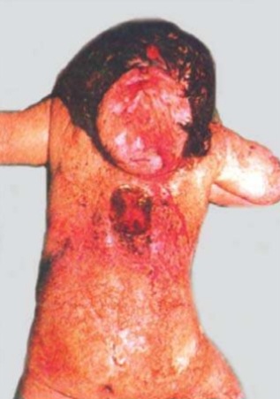
❓ Q1. Describe the findings in the image. What is the most likely diagnosis?.
✅ Model Answer:
• Findings: Midline occipital swelling, skin-covered, cystic, non-pulsatile, in posterior fontanelle/occipital region.
• Diagnosis: Occipital encephalocele – neural tube defect with herniation of meninges and brain tissue through skull defect.
• Difference from meningocele: Encephalocele contains neural tissue; meningocele contains only meninges/CSF, no neural tissue, normal neurology.
• Findings: Midline occipital swelling, skin-covered, cystic, non-pulsatile, in posterior fontanelle/occipital region.
• Diagnosis: Occipital encephalocele – neural tube defect with herniation of meninges and brain tissue through skull defect.
• Difference from meningocele: Encephalocele contains neural tissue; meningocele contains only meninges/CSF, no neural tissue, normal neurology.
❓ Q2. What is the embryological basis of occipital encephalocele? What are the risk factors?
✅ Model Answer:
• Embryology: Failure of closure of anterior (cranial) neural tube during 3rd-4th week of gestation (neurulation).
• Risk factors: Maternal folic acid deficiency (periconceptional), maternal diabetes, obesity, hyperthermia, valproate/carbamazepine use, family history (recurrence risk 3-5%).
• Prevention: Periconceptional folic acid (400 mcg/day routine; 4 mg/day for high-risk mothers).
• Occipital location most common in Western populations (~75%).
• Embryology: Failure of closure of anterior (cranial) neural tube during 3rd-4th week of gestation (neurulation).
• Risk factors: Maternal folic acid deficiency (periconceptional), maternal diabetes, obesity, hyperthermia, valproate/carbamazepine use, family history (recurrence risk 3-5%).
• Prevention: Periconceptional folic acid (400 mcg/day routine; 4 mg/day for high-risk mothers).
• Occipital location most common in Western populations (~75%).
❓ Q3. What are the common associated anomalies in occipital encephalocele?
✅ Model Answer:
• CNS: Hydrocephalus (50-70%), Chiari III malformation, corpus callosum agenesis, cerebellar hypoplasia, Dandy-Walker, seizures.
• Systemic: Cardiac defects, renal anomalies, Meckel-Gruber syndrome (encephalocele + polycystic kidneys + polydactyly – lethal), Walker-Warburg syndrome, trisomy 13/18.
• Important: Urgent MRI to evaluate hydrocephalus and other brain anomalies.
• CNS: Hydrocephalus (50-70%), Chiari III malformation, corpus callosum agenesis, cerebellar hypoplasia, Dandy-Walker, seizures.
• Systemic: Cardiac defects, renal anomalies, Meckel-Gruber syndrome (encephalocele + polycystic kidneys + polydactyly – lethal), Walker-Warburg syndrome, trisomy 13/18.
• Important: Urgent MRI to evaluate hydrocephalus and other brain anomalies.
❓ Q4. What are the differential diagnoses of a midline occipital swelling in a newborn?
✅ Model Answer:
• Occipital encephalocele (neural tissue, midline, bony defect)
• Cephalohematoma (subperiosteal, does not cross sutures, non-midline)
• Caput succedaneum (diffuse edema, crosses sutures, resolves days)
• Cystic hygroma (lateral/posterolateral, multiloculated, transilluminates)
• Dermoid cyst (midline, firm, non-transilluminating)
• Subgaleal hematoma (fluctuant, crosses sutures, traumatic)
• Occipital meningocele (only meninges/CSF, normal neurology)
• Key differentiating investigation: MRI to identify neural tissue and skull defect.
• Occipital encephalocele (neural tissue, midline, bony defect)
• Cephalohematoma (subperiosteal, does not cross sutures, non-midline)
• Caput succedaneum (diffuse edema, crosses sutures, resolves days)
• Cystic hygroma (lateral/posterolateral, multiloculated, transilluminates)
• Dermoid cyst (midline, firm, non-transilluminating)
• Subgaleal hematoma (fluctuant, crosses sutures, traumatic)
• Occipital meningocele (only meninges/CSF, normal neurology)
• Key differentiating investigation: MRI to identify neural tissue and skull defect.
❓ Q5. What is your immediate management for this infant in the newborn period?
✅ Model Answer:
1. Protect sac: cover with sterile, saline-moistened non-adherent dressing. Avoid pressure/rupture.
2. Positioning: side-lying to avoid pressure on sac.
3. Neurological assessment: baseline tone, reflexes, suck, limb movement.
4. Head circumference monitoring: daily for hydrocephalus.
5. Head ultrasound: bedside assessment of ventriculomegaly.
6. MRI brain/spine: definitive imaging for sac contents, brain anomalies, hydrocephalus.
7. Echocardiogram and renal ultrasound: screen associated anomalies.
8. Prophylactic antibiotics: if sac compromised or before repair.
9. Neurosurgery consultation: urgent for surgical planning.
1. Protect sac: cover with sterile, saline-moistened non-adherent dressing. Avoid pressure/rupture.
2. Positioning: side-lying to avoid pressure on sac.
3. Neurological assessment: baseline tone, reflexes, suck, limb movement.
4. Head circumference monitoring: daily for hydrocephalus.
5. Head ultrasound: bedside assessment of ventriculomegaly.
6. MRI brain/spine: definitive imaging for sac contents, brain anomalies, hydrocephalus.
7. Echocardiogram and renal ultrasound: screen associated anomalies.
8. Prophylactic antibiotics: if sac compromised or before repair.
9. Neurosurgery consultation: urgent for surgical planning.
❓ Q6. When is surgical repair performed? What are the surgical principles?
✅ Model Answer:
• Timing: Within first few days to weeks (elective early). Earlier if sac ruptured or leaking (emergency).
• Principles: Resection of sac, preservation of viable neural tissue (return to intracranial space if possible), watertight dural closure, closure of skull defect, skin closure (may need flaps/graft).
• Concurrent: VP shunt for hydrocephalus (same time or staged).
• Timing: Within first few days to weeks (elective early). Earlier if sac ruptured or leaking (emergency).
• Principles: Resection of sac, preservation of viable neural tissue (return to intracranial space if possible), watertight dural closure, closure of skull defect, skin closure (may need flaps/graft).
• Concurrent: VP shunt for hydrocephalus (same time or staged).
❓ Q7. How do you diagnose and manage hydrocephalus associated with occipital encephalocele?
✅ Model Answer:
• Diagnosis: Rapid head circumference growth, bulging fontanel, sunset sign. Cranial ultrasound/MRI showing enlarged ventricles (ventricular index >97th percentile).
• Incidence: 50-70% of occipital encephalocele patients.
• Management: Mild/stable – observe. Progressive/symptomatic – VP shunt insertion (may be before, same time, or after encephalocele repair). ETV alternative. Shunt complications: infection, obstruction, revision (~40% within first year).
• Diagnosis: Rapid head circumference growth, bulging fontanel, sunset sign. Cranial ultrasound/MRI showing enlarged ventricles (ventricular index >97th percentile).
• Incidence: 50-70% of occipital encephalocele patients.
• Management: Mild/stable – observe. Progressive/symptomatic – VP shunt insertion (may be before, same time, or after encephalocele repair). ETV alternative. Shunt complications: infection, obstruction, revision (~40% within first year).
❓ Q8. What is the neurological prognosis for an infant with an occipital encephalocele?
✅ Model Answer:
• Depends on: Sac content (neural tissue vs meninges only), volume of herniated brain, associated brain anomalies, hydrocephalus.
• Meningocele (no neural tissue): excellent prognosis, normal IQ.
• Encephalocele: Excellent (30-40% normal), moderate impairment (30-40% learning disabilities, mild motor deficits), severe impairment (20-30% intellectual disability, spastic quadriparesis, epilepsy).
• Mortality: 10-20% (higher with severe associated anomalies or Meckel-Gruber).
• Depends on: Sac content (neural tissue vs meninges only), volume of herniated brain, associated brain anomalies, hydrocephalus.
• Meningocele (no neural tissue): excellent prognosis, normal IQ.
• Encephalocele: Excellent (30-40% normal), moderate impairment (30-40% learning disabilities, mild motor deficits), severe impairment (20-30% intellectual disability, spastic quadriparesis, epilepsy).
• Mortality: 10-20% (higher with severe associated anomalies or Meckel-Gruber).
❓ Q9. Can occipital encephalocele be diagnosed prenatally? What are the sonographic findings?
✅ Model Answer:
• Yes, commonly diagnosed prenatally.
• Ultrasound findings: Midline cystic or solid-cystic occipital mass, skull defect, "lemon sign" (bifrontal indentation), "banana sign" (cerebellar herniation), hydrocephalus, associated anomalies (polycystic kidneys, polydactyly in Meckel-Gruber).
• Fetal MRI: Better delineation of sac contents and brain anomalies.
• Elevated maternal serum AFP and amniotic fluid AFP/AChE.
• Genetic testing: Amniocentesis for karyotype/microarray (trisomy 13,18, Meckel-Gruber genes).
• Yes, commonly diagnosed prenatally.
• Ultrasound findings: Midline cystic or solid-cystic occipital mass, skull defect, "lemon sign" (bifrontal indentation), "banana sign" (cerebellar herniation), hydrocephalus, associated anomalies (polycystic kidneys, polydactyly in Meckel-Gruber).
• Fetal MRI: Better delineation of sac contents and brain anomalies.
• Elevated maternal serum AFP and amniotic fluid AFP/AChE.
• Genetic testing: Amniocentesis for karyotype/microarray (trisomy 13,18, Meckel-Gruber genes).
❓ Q10. What genetic syndromes are associated with occipital encephalocele? Name two with their key features.
✅ Model Answer:
1. Meckel-Gruber syndrome (autosomal recessive): Triad – occipital encephalocele + polycystic kidneys + postaxial polydactyly. Also hepatic fibrosis, cardiac defects. Lethal.
2. Walker-Warburg syndrome (autosomal recessive): Encephalocele, hydrocephalus, cobblestone lissencephaly, cerebellar hypoplasia, congenital muscular dystrophy, eye anomalies (retinal dysplasia, cataracts). Severe neurodevelopmental impairment; early death.
Others: Joubert syndrome, Knobloch syndrome, trisomy 13/18.
1. Meckel-Gruber syndrome (autosomal recessive): Triad – occipital encephalocele + polycystic kidneys + postaxial polydactyly. Also hepatic fibrosis, cardiac defects. Lethal.
2. Walker-Warburg syndrome (autosomal recessive): Encephalocele, hydrocephalus, cobblestone lissencephaly, cerebellar hypoplasia, congenital muscular dystrophy, eye anomalies (retinal dysplasia, cataracts). Severe neurodevelopmental impairment; early death.
Others: Joubert syndrome, Knobloch syndrome, trisomy 13/18.
❓ Q11. What is the role of folic acid in preventing occipital encephalocele? What dose is recommended for recurrence prevention?
✅ Model Answer:
• Folic acid reduces NTD risk by ~70% when taken periconceptually.
• General prevention: 400 mcg (0.4 mg) daily.
• High-risk (previous NTD-affected pregnancy, maternal anticonvulsants, family history): 4 mg (4000 mcg) daily starting 1 month before conception through first trimester.
• Note: Not all NTDs preventable by folic acid (genetic causes like Meckel-Gruber) – need genetic counseling.
• Folic acid reduces NTD risk by ~70% when taken periconceptually.
• General prevention: 400 mcg (0.4 mg) daily.
• High-risk (previous NTD-affected pregnancy, maternal anticonvulsants, family history): 4 mg (4000 mcg) daily starting 1 month before conception through first trimester.
• Note: Not all NTDs preventable by folic acid (genetic causes like Meckel-Gruber) – need genetic counseling.
❓ Q12. How will you counsel the parents about the diagnosis and prognosis?
✅ Model Answer:
• “Your baby has a birth defect where the skull didn’t close completely, and a sac containing brain tissue and fluid has pushed out at the back of the head.”
• “We will protect the sac, do an MRI, and plan surgery to put brain tissue back and close the defect.”
• “Outcome depends on how much brain tissue is in the sac and other brain abnormalities. Some children develop normally; others may have learning or physical difficulties.”
• “Your baby may need a shunt for fluid on the brain (hydrocephalus).”
• “If isolated (non-syndromic), recurrence risk for future siblings is 3-5%. You should take high-dose folic acid (4 mg daily) before any future pregnancy.”
• “Nothing you did caused this. Folic acid reduces risk, but sometimes it happens despite best prevention.”
• “Your baby has a birth defect where the skull didn’t close completely, and a sac containing brain tissue and fluid has pushed out at the back of the head.”
• “We will protect the sac, do an MRI, and plan surgery to put brain tissue back and close the defect.”
• “Outcome depends on how much brain tissue is in the sac and other brain abnormalities. Some children develop normally; others may have learning or physical difficulties.”
• “Your baby may need a shunt for fluid on the brain (hydrocephalus).”
• “If isolated (non-syndromic), recurrence risk for future siblings is 3-5%. You should take high-dose folic acid (4 mg daily) before any future pregnancy.”
• “Nothing you did caused this. Folic acid reduces risk, but sometimes it happens despite best prevention.”
📋 Mock Test Feedback
💡 Examiner's note: Compare your answers with the model answers. In real TOACS, you would discuss these with the examiner.