⚕️ FCPS MCPS IMM MD Paediatrics TOACS · Mock Test
· 8-Minute Observed Station
📚 paeds.online
⏱️ TIME REMAINING
08:00
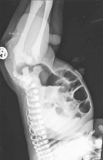
❓ Q1. Describe the radiographic findings. What is this study called and what is its purpose?
✅ Model Answer:
• Study: Invertogram (prone cross-table lateral x-ray).
• Positioning: Prone position with pelvis elevated (head down) – the "invertogram" view.
• Timing: Performed at 24 hours of age (allows air to reach the distal rectum).
• Purpose: To determine the level of the anorectal malformation (high vs low lesion) by measuring the distance between the distal rectal gas shadow and the perineal skin.
• Key finding: Air-filled distal rectum visible as a gas shadow.
• Study: Invertogram (prone cross-table lateral x-ray).
• Positioning: Prone position with pelvis elevated (head down) – the "invertogram" view.
• Timing: Performed at 24 hours of age (allows air to reach the distal rectum).
• Purpose: To determine the level of the anorectal malformation (high vs low lesion) by measuring the distance between the distal rectal gas shadow and the perineal skin.
• Key finding: Air-filled distal rectum visible as a gas shadow.
❓ Q2. How do you interpret the invertogram? What is the significance of the distance measured?
✅ Model Answer:
• The distance between the distal rectal gas (most inferior aspect) and the perineal skin is measured.
• Low lesion: Rectal gas < 1 cm from the perineal skin → primary perineal anoplasty can be performed.
• High lesion: Rectal gas ≥ 1 cm from the perineal skin → requires a protective colostomy (since the rectum is too high for a primary perineal approach).
• The 1 cm threshold is a key surgical decision point.
• The distance between the distal rectal gas (most inferior aspect) and the perineal skin is measured.
• Low lesion: Rectal gas < 1 cm from the perineal skin → primary perineal anoplasty can be performed.
• High lesion: Rectal gas ≥ 1 cm from the perineal skin → requires a protective colostomy (since the rectum is too high for a primary perineal approach).
• The 1 cm threshold is a key surgical decision point.
❓ Q3. Why is the invertogram performed at 24 hours of life, not immediately after birth?
✅ Model Answer:
• The invertogram is performed at 24 hours of age to allow swallowed air to reach the distal rectum.
• In the first 12-18 hours, air may not have passed through the entire gastrointestinal tract, so the rectal gas shadow may not be visible.
• Waiting 24 hours ensures that the distal rectum is adequately distended with air for accurate measurement.
• If the infant has a high lesion or a fistula, air may escape through the fistula, affecting the measurement.
• The invertogram is performed at 24 hours of age to allow swallowed air to reach the distal rectum.
• In the first 12-18 hours, air may not have passed through the entire gastrointestinal tract, so the rectal gas shadow may not be visible.
• Waiting 24 hours ensures that the distal rectum is adequately distended with air for accurate measurement.
• If the infant has a high lesion or a fistula, air may escape through the fistula, affecting the measurement.
❓ Q4. What are the common types of anorectal malformations in males and females?
✅ Model Answer:
• In males (most common):
- Rectourethral fistula (bulbar or prostatic) – most common high lesion.
- Rectovesical fistula – rare, high lesion.
- Perineal fistula – low lesion (rectum opens to perineum).
- Anal stenosis – low lesion.
- Imperforate anus without fistula – rare.
• In females (most common):
- Rectovestibular fistula – most common (fistula to the vestibule of the vagina).
- Perineal fistula – low lesion.
- Rectovaginal fistula – less common.
- Cloacal malformation – complex (rectum, vagina, and urethra join to a common channel).
• In males (most common):
- Rectourethral fistula (bulbar or prostatic) – most common high lesion.
- Rectovesical fistula – rare, high lesion.
- Perineal fistula – low lesion (rectum opens to perineum).
- Anal stenosis – low lesion.
- Imperforate anus without fistula – rare.
• In females (most common):
- Rectovestibular fistula – most common (fistula to the vestibule of the vagina).
- Perineal fistula – low lesion.
- Rectovaginal fistula – less common.
- Cloacal malformation – complex (rectum, vagina, and urethra join to a common channel).
❓ Q5. What is the VACTERL association? Why is it important in anorectal malformations?
✅ Model Answer:
• VACTERL association: Vertebral anomalies, Anal atresia, Cardiac defects, TE fistula, Esophageal atresia, Renal anomalies, Limb anomalies.
• Importance: Anorectal malformations are frequently associated with other anomalies in the VACTERL spectrum.
• Screening: All neonates with anorectal malformations should have:
- Echocardiogram (cardiac anomalies).
- Renal ultrasound (renal anomalies).
- Spinal ultrasound / MRI (vertebral/sacral anomalies).
- X-ray of the spine (sacral anomalies – hemisacrum, sacral agenesis).
• VACTERL association: Vertebral anomalies, Anal atresia, Cardiac defects, TE fistula, Esophageal atresia, Renal anomalies, Limb anomalies.
• Importance: Anorectal malformations are frequently associated with other anomalies in the VACTERL spectrum.
• Screening: All neonates with anorectal malformations should have:
- Echocardiogram (cardiac anomalies).
- Renal ultrasound (renal anomalies).
- Spinal ultrasound / MRI (vertebral/sacral anomalies).
- X-ray of the spine (sacral anomalies – hemisacrum, sacral agenesis).
❓ Q6. A newborn with a high anorectal malformation (rectal gas ≥1 cm from perineum). What is the surgical management?
✅ Model Answer:
• Neonatal management:
1. Protective colostomy – usually a divided sigmoid colostomy (to divert stool and prevent the infant from passing meconium through a fistula).
2. This allows the infant to be fed and grow before definitive repair.
3. Careful monitoring for urinary retention or sepsis (if a rectourethral fistula is present).
• Definitive repair (3-6 months):
- PSARP (Posterior Sagittal Anorectoplasty – Pena procedure) – the gold standard.
- The rectum is dissected and brought down to the perineum through a posterior sagittal incision.
- The fistula is closed.
- The sphincter complex is preserved.
• After PSARP: The colostomy is closed 4-6 weeks later.
• Neonatal management:
1. Protective colostomy – usually a divided sigmoid colostomy (to divert stool and prevent the infant from passing meconium through a fistula).
2. This allows the infant to be fed and grow before definitive repair.
3. Careful monitoring for urinary retention or sepsis (if a rectourethral fistula is present).
• Definitive repair (3-6 months):
- PSARP (Posterior Sagittal Anorectoplasty – Pena procedure) – the gold standard.
- The rectum is dissected and brought down to the perineum through a posterior sagittal incision.
- The fistula is closed.
- The sphincter complex is preserved.
• After PSARP: The colostomy is closed 4-6 weeks later.
❓ Q7. What is the Pena procedure (PSARP)? When is it indicated?
✅ Model Answer:
• PSARP: Posterior Sagittal Anorectoplasty (Pena procedure).
• Indications: High anorectal malformations (rectal gas ≥1 cm from perineum) and intermediate lesions.
• Procedure:
- A midline posterior sagittal incision is made from the coccyx to the perineum.
- The rectum and fistula are identified.
- The fistula is closed.
- The rectum is mobilized and brought down to the perineum.
- The rectum is sutured to the anal sphincter complex.
• Advantages:
- Excellent visualization of the anatomy.
- Good preservation of the sphincter complex.
- Good functional outcomes (continence).
• PSARP: Posterior Sagittal Anorectoplasty (Pena procedure).
• Indications: High anorectal malformations (rectal gas ≥1 cm from perineum) and intermediate lesions.
• Procedure:
- A midline posterior sagittal incision is made from the coccyx to the perineum.
- The rectum and fistula are identified.
- The fistula is closed.
- The rectum is mobilized and brought down to the perineum.
- The rectum is sutured to the anal sphincter complex.
• Advantages:
- Excellent visualization of the anatomy.
- Good preservation of the sphincter complex.
- Good functional outcomes (continence).
❓ Q8. A newborn with anorectal malformation and a perineal fistula (low lesion). What is the surgical management?
✅ Model Answer:
• Primary perineal anoplasty – the definitive procedure can be performed in the neonatal period.
• Procedure:
- The fistula is identified on the perineum.
- The rectum is mobilized through the perineal approach.
- The rectum is brought down and sutured to the perineal skin.
- No colostomy is required.
• Advantages:
- Single-stage repair.
- No colostomy morbidity.
- Excellent continence outcomes (~90%).
• Primary perineal anoplasty – the definitive procedure can be performed in the neonatal period.
• Procedure:
- The fistula is identified on the perineum.
- The rectum is mobilized through the perineal approach.
- The rectum is brought down and sutured to the perineal skin.
- No colostomy is required.
• Advantages:
- Single-stage repair.
- No colostomy morbidity.
- Excellent continence outcomes (~90%).
❓ Q9. What is the Currarino triad? How does it relate to anorectal malformations?
✅ Model Answer:
• Currarino triad: A rare condition consisting of:
1. Anorectal malformation (usually anal stenosis or imperforate anus).
2. Sacral anomaly (hemisacrum, sacral agenesis, or scimitar sacrum).
3. Presacral mass (teratoma, meningocele, or enteric cyst).
• Important: The presacral mass can cause constipation, urinary retention, or bowel obstruction.
• Diagnosis: MRI of the spine and pelvis.
• Management: Surgical excision of the presacral mass + repair of the anorectal malformation.
• Currarino triad: A rare condition consisting of:
1. Anorectal malformation (usually anal stenosis or imperforate anus).
2. Sacral anomaly (hemisacrum, sacral agenesis, or scimitar sacrum).
3. Presacral mass (teratoma, meningocele, or enteric cyst).
• Important: The presacral mass can cause constipation, urinary retention, or bowel obstruction.
• Diagnosis: MRI of the spine and pelvis.
• Management: Surgical excision of the presacral mass + repair of the anorectal malformation.
❓ Q10. What is the role of ultrasound or MRI in anorectal malformations?
✅ Model Answer:
• Spinal ultrasound: To screen for spinal cord anomalies (tethered cord, lipomeningocele, sacral agenesis) – performed in the neonatal period.
• MRI of the spine and pelvis: More detailed assessment of sacral anomalies, presacral masses (Currarino triad), and the sphincter complex.
• Renal ultrasound: To screen for renal anomalies (hydronephrosis, renal agenesis, cystic kidneys).
• Echocardiogram: To screen for cardiac anomalies (VACTERL).
• Distal colostogram: Performed after colostomy to define the anatomy of the distal rectum and fistula before PSARP.
• Spinal ultrasound: To screen for spinal cord anomalies (tethered cord, lipomeningocele, sacral agenesis) – performed in the neonatal period.
• MRI of the spine and pelvis: More detailed assessment of sacral anomalies, presacral masses (Currarino triad), and the sphincter complex.
• Renal ultrasound: To screen for renal anomalies (hydronephrosis, renal agenesis, cystic kidneys).
• Echocardiogram: To screen for cardiac anomalies (VACTERL).
• Distal colostogram: Performed after colostomy to define the anatomy of the distal rectum and fistula before PSARP.
❓ Q11. What is the long-term prognosis for children with repaired anorectal malformations?
✅ Model Answer:
• Low lesions (perineal fistula): Excellent prognosis – 90% have normal continence.
• High lesions (rectourethral fistula): Variable prognosis – 40-60% have good continence. May require a bowel management program (enemas, laxatives).
• Factors affecting prognosis:
- Level of the lesion (higher = poorer continence).
- Associated anomalies (sacral agenesis, spinal cord anomalies).
- Quality of the sphincter complex.
- Surgical technique (PSARP gives better outcomes).
- Post-operative bowel management.
• Long-term follow-up: Lifelong monitoring for constipation, soiling, and urinary function.
• Low lesions (perineal fistula): Excellent prognosis – 90% have normal continence.
• High lesions (rectourethral fistula): Variable prognosis – 40-60% have good continence. May require a bowel management program (enemas, laxatives).
• Factors affecting prognosis:
- Level of the lesion (higher = poorer continence).
- Associated anomalies (sacral agenesis, spinal cord anomalies).
- Quality of the sphincter complex.
- Surgical technique (PSARP gives better outcomes).
- Post-operative bowel management.
• Long-term follow-up: Lifelong monitoring for constipation, soiling, and urinary function.
❓ Q12. A newborn with anorectal malformation has a flat perineum and no visible fistula. The invertogram shows rectal gas ≥1 cm from the perineum. What is the next step in management?
✅ Model Answer:
• This is a high anorectal malformation (likely rectourethral fistula in a male).
• Next steps:
1. Protective colostomy – a divided sigmoid colostomy (to divert stool).
2. Urgent ultrasound – renal ultrasound to check for hydronephrosis (risk of urinary retention from the fistula).
3. Echocardiogram – to screen for cardiac anomalies (VACTERL).
4. Spinal ultrasound – to screen for spinal anomalies.
5. Definitive repair (PSARP) – at 3-6 months of age after the colostomy.
6. Distal colostogram – before PSARP to delineate the anatomy.
• This is a high anorectal malformation (likely rectourethral fistula in a male).
• Next steps:
1. Protective colostomy – a divided sigmoid colostomy (to divert stool).
2. Urgent ultrasound – renal ultrasound to check for hydronephrosis (risk of urinary retention from the fistula).
3. Echocardiogram – to screen for cardiac anomalies (VACTERL).
4. Spinal ultrasound – to screen for spinal anomalies.
5. Definitive repair (PSARP) – at 3-6 months of age after the colostomy.
6. Distal colostogram – before PSARP to delineate the anatomy.
⚠️ Key concept: The invertogram (prone cross‑table lateral x‑ray) determines the level of the anorectal malformation.
Rectal gas ≥1 cm from perineal skin indicates a high lesion → needs a protective colostomy
followed by PSARP at 3‑6 months.
Gas <1 cm = low lesion → primary perineal anoplasty.
📋 Mock Test Feedback
💡 Examiner's note: Compare your answers with the model answers. In real TOACS, you would discuss these with the examiner.