⚕️ FCPS MCPS IMM MD Paediatrics TOACS · Mock Test
X-ray Long Bones · 8-Minute Observed Station
📚 paeds.online
⏱️ TIME REMAINING
08:00
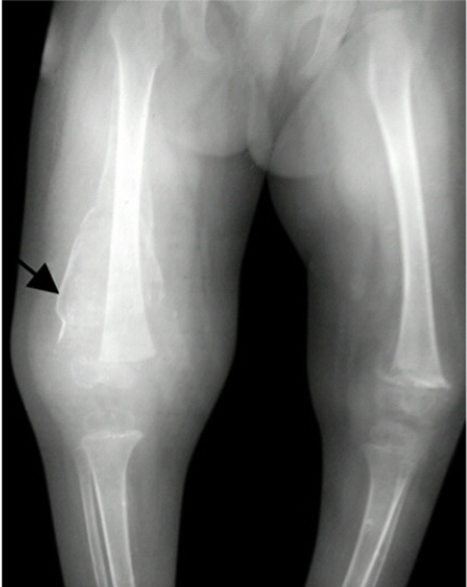
3 Yrs old child with Failure to thrive now c/o refusal to walk and leg pain for 2 weeks.
❓ Q1. Describe the radiographic findings. What is the most likely diagnosis?
✅ Model Answer:
• Frankel line – dense white line at the metaphysis (thickened zone of provisional calcification).
• Trummerfeld zone – lucent band of rarefaction immediately below the Frankel line.
• Pelkan spur – lateral prolongation of the white line at the cortical ends.
• Wimberger ring sign – dense thin line outlining the epiphyseal center.
• Subperiosteal hemorrhage (periosteal elevation).
• Diagnosis: Scurvy (vitamin C deficiency).
• Frankel line – dense white line at the metaphysis (thickened zone of provisional calcification).
• Trummerfeld zone – lucent band of rarefaction immediately below the Frankel line.
• Pelkan spur – lateral prolongation of the white line at the cortical ends.
• Wimberger ring sign – dense thin line outlining the epiphyseal center.
• Subperiosteal hemorrhage (periosteal elevation).
• Diagnosis: Scurvy (vitamin C deficiency).
❓ Q2. What is the Frankel line and why does it occur?
✅ Model Answer:
• Frankel line: A dense, white line seen at the metaphysis of long bones in scurvy.
• Mechanism: It represents a thickened zone of provisional calcification – due to impaired collagen synthesis (vitamin C deficiency), osteoblasts fail to form osteoid, but calcification continues at the primary spongiosa.
• The line is pathognomonic for scurvy.
• It is the dense calcium deposited at the metaphyseal growth plate.
• It may be seen in both the distal femur and proximal tibia.
• Frankel line: A dense, white line seen at the metaphysis of long bones in scurvy.
• Mechanism: It represents a thickened zone of provisional calcification – due to impaired collagen synthesis (vitamin C deficiency), osteoblasts fail to form osteoid, but calcification continues at the primary spongiosa.
• The line is pathognomonic for scurvy.
• It is the dense calcium deposited at the metaphyseal growth plate.
• It may be seen in both the distal femur and proximal tibia.
❓ Q3. What is the Trummerfeld zone? What does it indicate?
✅ Model Answer:
• Trummerfeld zone: A lucent (radiolucent) band of rarefaction immediately below the Frankel line.
• Mechanism: It represents fragile, osteopenic bone in the metaphysis due to impaired osteoid formation and trabecular bone resorption.
• The combination of the dense Frankel line above and the lucent Trummerfeld zone below is pathognomonic for scurvy.
• Also called the "scorbutic line" or "zone of rarefaction".
• It indicates active vitamin C deficiency and impaired bone matrix formation.
• Trummerfeld zone: A lucent (radiolucent) band of rarefaction immediately below the Frankel line.
• Mechanism: It represents fragile, osteopenic bone in the metaphysis due to impaired osteoid formation and trabecular bone resorption.
• The combination of the dense Frankel line above and the lucent Trummerfeld zone below is pathognomonic for scurvy.
• Also called the "scorbutic line" or "zone of rarefaction".
• It indicates active vitamin C deficiency and impaired bone matrix formation.
❓ Q4. What is the Pelkan spur and what does it indicate?
✅ Model Answer:
• Pelkan spur: A lateral prolongation or "spur" of the Frankel line at the cortical ends of the metaphysis.
• Mechanism: The dense calcified cartilage extends laterally beyond the cortical margin.
• Significance: It is a classic radiographic sign of scurvy.
• May be seen as a small "spike" or "spur" extending from the metaphyseal edges.
• Helps differentiate scurvy from other causes of metaphyseal dysplasia.
• Pelkan spur: A lateral prolongation or "spur" of the Frankel line at the cortical ends of the metaphysis.
• Mechanism: The dense calcified cartilage extends laterally beyond the cortical margin.
• Significance: It is a classic radiographic sign of scurvy.
• May be seen as a small "spike" or "spur" extending from the metaphyseal edges.
• Helps differentiate scurvy from other causes of metaphyseal dysplasia.
❓ Q5. What is the Wimberger ring sign? Where is it seen?
✅ Model Answer:
• Wimberger ring sign: A dense, thin line outlining the epiphyseal center (the ossification center of the epiphysis).
• Location: Most commonly seen in the distal femoral epiphysis.
• Mechanism: The epiphyseal ossification center is outlined by a dense calcified margin, while the central portion may be radiolucent.
• Significance: It is a classic radiographic sign of scurvy.
• It may also be seen in the proximal tibial epiphysis.
• The sign represents the dense calcified ring around the epiphyseal center.
• Wimberger ring sign: A dense, thin line outlining the epiphyseal center (the ossification center of the epiphysis).
• Location: Most commonly seen in the distal femoral epiphysis.
• Mechanism: The epiphyseal ossification center is outlined by a dense calcified margin, while the central portion may be radiolucent.
• Significance: It is a classic radiographic sign of scurvy.
• It may also be seen in the proximal tibial epiphysis.
• The sign represents the dense calcified ring around the epiphyseal center.
❓ Q6. What are the clinical features of scurvy in children?
✅ Model Answer:
• Pseudoparalysis – refusal to walk/use limbs due to pain (subperiosteal hemorrhage).
• Leg pain and tenderness – along the long bones.
• Swollen, bleeding gums – gingival hypertrophy, bleeding, petechiae.
• Perifollicular hemorrhage – petechiae or bruising around hair follicles.
• Ecchymoses – easy bruising.
• Anemia – normocytic/normochromic (due to impaired iron absorption and bleeding).
• Irritability and lethargy.
• Poor wound healing.
• Costochondral beading – "scorbutic rosary" (prominent costochondral junctions).
• Pseudoparalysis – refusal to walk/use limbs due to pain (subperiosteal hemorrhage).
• Leg pain and tenderness – along the long bones.
• Swollen, bleeding gums – gingival hypertrophy, bleeding, petechiae.
• Perifollicular hemorrhage – petechiae or bruising around hair follicles.
• Ecchymoses – easy bruising.
• Anemia – normocytic/normochromic (due to impaired iron absorption and bleeding).
• Irritability and lethargy.
• Poor wound healing.
• Costochondral beading – "scorbutic rosary" (prominent costochondral junctions).
❓ Q7. What is the pathophysiology of scurvy? Why does vitamin C deficiency cause these changes?
✅ Model Answer:
• Vitamin C (ascorbic acid) is a cofactor for hydroxylation of proline and lysine – required for collagen synthesis.
• Deficiency leads to impaired collagen synthesis.
• Effects:
- Fragile capillaries → petechiae, purpura, subperiosteal hemorrhage.
- Impaired osteoid formation → defective bone matrix → fragility.
- Impaired wound healing.
- Weak connective tissue → bleeding gums, perifollicular hemorrhage.
- Bone changes: The Frankel line represents calcified cartilage at the metaphysis; the Trummerfeld zone is the fragile, osteopenic bone underneath.
• Vitamin C (ascorbic acid) is a cofactor for hydroxylation of proline and lysine – required for collagen synthesis.
• Deficiency leads to impaired collagen synthesis.
• Effects:
- Fragile capillaries → petechiae, purpura, subperiosteal hemorrhage.
- Impaired osteoid formation → defective bone matrix → fragility.
- Impaired wound healing.
- Weak connective tissue → bleeding gums, perifollicular hemorrhage.
- Bone changes: The Frankel line represents calcified cartilage at the metaphysis; the Trummerfeld zone is the fragile, osteopenic bone underneath.
❓ Q8. What are the risk factors for developing scurvy in children?
✅ Model Answer:
• Restricted diets – autism spectrum disorder, severe food selectivity.
• Poverty – limited access to fresh fruits and vegetables.
• Malabsorption – Crohn's disease, cystic fibrosis, celiac disease.
• Chronic illness – malignancy, renal disease.
• Exclusive formula feeding without vitamin C supplementation (rare).
• Eating disorders – anorexia nervosa.
• Breastfed infants of vitamin C-deficient mothers.
• Fad diets – avoiding fruits and vegetables.
• Restricted diets – autism spectrum disorder, severe food selectivity.
• Poverty – limited access to fresh fruits and vegetables.
• Malabsorption – Crohn's disease, cystic fibrosis, celiac disease.
• Chronic illness – malignancy, renal disease.
• Exclusive formula feeding without vitamin C supplementation (rare).
• Eating disorders – anorexia nervosa.
• Breastfed infants of vitamin C-deficient mothers.
• Fad diets – avoiding fruits and vegetables.
❓ Q9. How do you diagnose scurvy? What laboratory tests support the diagnosis?
✅ Model Answer:
• Diagnosis: Clinical presentation + characteristic X-ray findings (Frankel line, Trummerfeld zone, Pelkan spur).
• Laboratory tests:
- Serum ascorbic acid (vitamin C) level – low (<11.4 µmol/L).
- Leukocyte ascorbic acid level – more reliable but less commonly used.
- Anemia – normocytic/normochromic, low serum iron (due to impaired iron absorption).
- Coagulation studies – normal (unlike vitamin K deficiency).
- Serum albumin – may be low in malnutrition.
• Response to treatment: Clinical improvement within 24-48 hours of vitamin C supplementation confirms the diagnosis.
• Diagnosis: Clinical presentation + characteristic X-ray findings (Frankel line, Trummerfeld zone, Pelkan spur).
• Laboratory tests:
- Serum ascorbic acid (vitamin C) level – low (<11.4 µmol/L).
- Leukocyte ascorbic acid level – more reliable but less commonly used.
- Anemia – normocytic/normochromic, low serum iron (due to impaired iron absorption).
- Coagulation studies – normal (unlike vitamin K deficiency).
- Serum albumin – may be low in malnutrition.
• Response to treatment: Clinical improvement within 24-48 hours of vitamin C supplementation confirms the diagnosis.
❓ Q10. What is the treatment for scurvy?
✅ Model Answer:
• Vitamin C (ascorbic acid) supplementation:
- 100-200 mg/day orally (divided doses).
- Duration: 2-3 months (until symptoms resolve).
- Rapid response: Clinical improvement within 24-48 hours (pain decreases, gums improve).
• Dietary modification:
- Increase intake of vitamin C-rich foods (citrus fruits, tomatoes, strawberries, kiwi, bell peppers, broccoli).
- Multi-vitamin supplementation.
• Supportive care:
- Pain management (analgesics).
- Dental care for gingival bleeding.
- Nutritional rehabilitation for underlying malnutrition.
• Follow-up: Monitor for clinical and radiological improvement (Frankel line resolves).
• Vitamin C (ascorbic acid) supplementation:
- 100-200 mg/day orally (divided doses).
- Duration: 2-3 months (until symptoms resolve).
- Rapid response: Clinical improvement within 24-48 hours (pain decreases, gums improve).
• Dietary modification:
- Increase intake of vitamin C-rich foods (citrus fruits, tomatoes, strawberries, kiwi, bell peppers, broccoli).
- Multi-vitamin supplementation.
• Supportive care:
- Pain management (analgesics).
- Dental care for gingival bleeding.
- Nutritional rehabilitation for underlying malnutrition.
• Follow-up: Monitor for clinical and radiological improvement (Frankel line resolves).
❓ Q11. How do you differentiate scurvy from rickets on X-ray?
✅ Model Answer:
• Scurvy:
- Frankel line (dense metaphyseal white line).
- Trummerfeld zone (lucent band below Frankel line).
- Pelkan spur (lateral spur).
- Wimberger ring (dense epiphyseal outline).
- Subperiosteal hemorrhage.
- No bowing or widening of the growth plate.
• Rickets:
- Widened, frayed, cupped metaphysis.
- No Frankel line (dense line is absent).
- Bowing of long bones.
- Looser zones (pseudofractures).
- No Wimberger ring sign.
- Key difference: Scurvy has a dense line at the metaphysis; rickets has a frayed, widened, cupped metaphysis.
• Scurvy:
- Frankel line (dense metaphyseal white line).
- Trummerfeld zone (lucent band below Frankel line).
- Pelkan spur (lateral spur).
- Wimberger ring (dense epiphyseal outline).
- Subperiosteal hemorrhage.
- No bowing or widening of the growth plate.
• Rickets:
- Widened, frayed, cupped metaphysis.
- No Frankel line (dense line is absent).
- Bowing of long bones.
- Looser zones (pseudofractures).
- No Wimberger ring sign.
- Key difference: Scurvy has a dense line at the metaphysis; rickets has a frayed, widened, cupped metaphysis.
❓ Q12. A child with autism and restricted diet presents with refusal to walk and leg pain. X-ray shows Frankel line, Trummerfeld zone, and Pelkan spur. What is the most likely diagnosis and what is the treatment?
✅ Model Answer:
• This is a classic presentation of scurvy (vitamin C deficiency).
• The triad of Frankel line, Trummerfeld zone, and Pelkan spur is pathognomonic for scurvy.
• Treatment:
- Vitamin C supplementation: 100-200 mg/day orally.
- Dietary modification: Increase vitamin C-rich foods.
- Pain management: Analgesics for leg pain.
- Follow-up: Clinical improvement within 24-48 hours; repeat X-ray in 2-3 months to monitor resolution.
• This is a classic presentation of scurvy (vitamin C deficiency).
• The triad of Frankel line, Trummerfeld zone, and Pelkan spur is pathognomonic for scurvy.
• Treatment:
- Vitamin C supplementation: 100-200 mg/day orally.
- Dietary modification: Increase vitamin C-rich foods.
- Pain management: Analgesics for leg pain.
- Follow-up: Clinical improvement within 24-48 hours; repeat X-ray in 2-3 months to monitor resolution.
⚠️ Key concept: The Frankel line (dense metaphyseal white line) with the Trummerfeld zone (lucent band underneath) is pathognomonic for scurvy.
Vitamin C deficiency causes impaired collagen synthesis, leading to fragile capillaries, subperiosteal hemorrhages, and characteristic radiographic changes.
📋 Mock Test Feedback
💡 Examiner's note: Compare your answers with the model answers. In real TOACS, you would discuss these with the examiner.