⚕️ FCPS MCPS IMM MD Paediatrics TOACS · Mock Test
X-ray Skull · 8-Minute Observed Station
📚 paeds.online
⏱️ TIME REMAINING
08:00
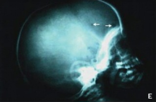
❓ Q1. Describe the radiographic findings. What is the most likely diagnosis?
✅ Model Answer:
• Lytic (punched-out) lesions – well-circumscribed, round/oval radiolucencies.
• Multiple lesions – often bilateral or scattered.
• Beveled edge – "hole-within-a-hole" appearance (classic sign).
• No sclerosis (unlike osteomyelitis or metastases).
• Intracranial extension may have soft tissue component.
• Diagnosis: Langerhans Cell Histiocytosis (LCH) – bone involvement (eosinophilic granuloma).
• Lytic (punched-out) lesions – well-circumscribed, round/oval radiolucencies.
• Multiple lesions – often bilateral or scattered.
• Beveled edge – "hole-within-a-hole" appearance (classic sign).
• No sclerosis (unlike osteomyelitis or metastases).
• Intracranial extension may have soft tissue component.
• Diagnosis: Langerhans Cell Histiocytosis (LCH) – bone involvement (eosinophilic granuloma).
❓ Q2. What is the "beveled edge" or "hole-within-a-hole" sign in LCH?
✅ Model Answer:
• Beveled edge sign: A radiographic appearance of skull lesions in LCH where the outer table is eroded more than the inner table, creating a "double contour" or "hole-within-a-hole" appearance.
• Mechanism: The lesions erode through the skull in a cone-like fashion, with the outer table eroded more extensively.
• It is a classic radiographic feature of Langerhans cell histiocytosis.
• Helps differentiate LCH from other lytic lesions (e.g., metastases, osteomyelitis).
• May be seen on both AP and lateral views.
• Beveled edge sign: A radiographic appearance of skull lesions in LCH where the outer table is eroded more than the inner table, creating a "double contour" or "hole-within-a-hole" appearance.
• Mechanism: The lesions erode through the skull in a cone-like fashion, with the outer table eroded more extensively.
• It is a classic radiographic feature of Langerhans cell histiocytosis.
• Helps differentiate LCH from other lytic lesions (e.g., metastases, osteomyelitis).
• May be seen on both AP and lateral views.
❓ Q3. What is the pathophysiology of Langerhans cell histiocytosis?
✅ Model Answer:
• LCH is a clonal proliferative disorder of Langerhans cells (CD1a+, langerin+).
• Pathogenesis: Activation of the MAPK pathway (RAS-RAF-MEK-ERK) – most commonly due to BRAF V600E mutation (present in ~50-60% of cases).
• Other mutations: MAP2K1, ARAF, NRAS, KRAS.
• The abnormal Langerhans cells infiltrate tissues and recruit inflammatory cells (eosinophils, lymphocytes), causing tissue destruction.
• This leads to the characteristic lytic bone lesions, skin rash, and organ infiltration.
• LCH is a clonal proliferative disorder of Langerhans cells (CD1a+, langerin+).
• Pathogenesis: Activation of the MAPK pathway (RAS-RAF-MEK-ERK) – most commonly due to BRAF V600E mutation (present in ~50-60% of cases).
• Other mutations: MAP2K1, ARAF, NRAS, KRAS.
• The abnormal Langerhans cells infiltrate tissues and recruit inflammatory cells (eosinophils, lymphocytes), causing tissue destruction.
• This leads to the characteristic lytic bone lesions, skin rash, and organ infiltration.
❓ Q4. What are the clinical presentations of LCH based on the involved organ systems?
✅ Model Answer:
• Single-system LCH:
- Bone (eosinophilic granuloma): Painful or painless swelling, lytic lesions (skull, femur, pelvis, spine).
- Skin: Seborrheic dermatitis, scaly erythematous rash (scalp, groin, postauricular).
- Lymph nodes.
• Multisystem LCH:
- Risk organs: Liver, spleen, bone marrow (indicates worse prognosis).
- Pituitary: Central diabetes insipidus (most common endocrine manifestation).
- Lung: Cystic lesions, pneumothorax.
- CNS: Hypothalamic-pituitary lesions, neurodegenerative changes.
- Ear: Chronic otitis media unresponsive to antibiotics.
- Oral: Gum swelling, tooth loss.
• Classic triad (Hand-Schüller-Christian): Lytic skull lesions, diabetes insipidus, exophthalmos.
• Single-system LCH:
- Bone (eosinophilic granuloma): Painful or painless swelling, lytic lesions (skull, femur, pelvis, spine).
- Skin: Seborrheic dermatitis, scaly erythematous rash (scalp, groin, postauricular).
- Lymph nodes.
• Multisystem LCH:
- Risk organs: Liver, spleen, bone marrow (indicates worse prognosis).
- Pituitary: Central diabetes insipidus (most common endocrine manifestation).
- Lung: Cystic lesions, pneumothorax.
- CNS: Hypothalamic-pituitary lesions, neurodegenerative changes.
- Ear: Chronic otitis media unresponsive to antibiotics.
- Oral: Gum swelling, tooth loss.
• Classic triad (Hand-Schüller-Christian): Lytic skull lesions, diabetes insipidus, exophthalmos.
❓ Q5. How is Langerhans cell histiocytosis diagnosed?
✅ Model Answer:
• Diagnosis is confirmed by biopsy of an affected site (bone, skin, lymph node).
• Histopathology:
- Langerhans cells (CD1a+, langerin+, S100+).
- Birbeck granules on electron microscopy (tennis-racket shape).
• Molecular testing: BRAF V600E mutation analysis (important for targeted therapy).
• Imaging:
- Skeletal survey (skull X-ray, long bones, spine, pelvis).
- MRI: Brain/pituitary for CNS involvement, spine for vertebral lesions.
- CT chest: For lung involvement.
• Laboratory:
- CBC (cytopenias indicate bone marrow involvement).
- Liver function tests (hepatomegaly/dysfunction).
- Urine osmolality (diabetes insipidus).
- Coagulation studies.
• Diagnosis is confirmed by biopsy of an affected site (bone, skin, lymph node).
• Histopathology:
- Langerhans cells (CD1a+, langerin+, S100+).
- Birbeck granules on electron microscopy (tennis-racket shape).
• Molecular testing: BRAF V600E mutation analysis (important for targeted therapy).
• Imaging:
- Skeletal survey (skull X-ray, long bones, spine, pelvis).
- MRI: Brain/pituitary for CNS involvement, spine for vertebral lesions.
- CT chest: For lung involvement.
• Laboratory:
- CBC (cytopenias indicate bone marrow involvement).
- Liver function tests (hepatomegaly/dysfunction).
- Urine osmolality (diabetes insipidus).
- Coagulation studies.
❓ Q6. How is Langerhans cell histiocytosis classified (single-system vs multisystem)?
✅ Model Answer:
• Single-system LCH:
- Involvement of one organ system (bone, skin, lymph node).
- Usually carries a good prognosis.
- May be unifocal (single lesion) or multifocal (multiple lesions in one system).
• Multisystem LCH:
- Involvement of two or more organ systems.
- Risk organs (RO+): Involvement of liver, spleen, or bone marrow → worse prognosis.
- Non-risk organs (RO-): Involvement of bone, skin, pituitary, lungs without risk organs.
• The classification determines treatment intensity and prognosis.
• Single-system LCH:
- Involvement of one organ system (bone, skin, lymph node).
- Usually carries a good prognosis.
- May be unifocal (single lesion) or multifocal (multiple lesions in one system).
• Multisystem LCH:
- Involvement of two or more organ systems.
- Risk organs (RO+): Involvement of liver, spleen, or bone marrow → worse prognosis.
- Non-risk organs (RO-): Involvement of bone, skin, pituitary, lungs without risk organs.
• The classification determines treatment intensity and prognosis.
❓ Q7. What is the treatment for Langerhans cell histiocytosis?
✅ Model Answer:
• Single-system, unifocal (bone):
- Observation, curettage, or local corticosteroid injection.
- No systemic therapy usually required.
• Single-system, multifocal:
- May require systemic therapy if extensive or symptomatic.
• Multisystem LCH (non-risk organs):
- Vinblastine + prednisone (6-12 months) – standard first-line.
• Multisystem LCH (risk organs involved):
- Vinblastine + prednisone + additional agents (e.g., mercaptopurine).
- Consider targeted therapy (BRAF inhibitors: vemurafenib, dabrafenib; MEK inhibitors: trametinib) if BRAF V600E-positive and refractory.
• Supportive care:
- Desmopressin (DDAVP) for diabetes insipidus.
- Pain management for bone lesions.
- Physical therapy.
• Single-system, unifocal (bone):
- Observation, curettage, or local corticosteroid injection.
- No systemic therapy usually required.
• Single-system, multifocal:
- May require systemic therapy if extensive or symptomatic.
• Multisystem LCH (non-risk organs):
- Vinblastine + prednisone (6-12 months) – standard first-line.
• Multisystem LCH (risk organs involved):
- Vinblastine + prednisone + additional agents (e.g., mercaptopurine).
- Consider targeted therapy (BRAF inhibitors: vemurafenib, dabrafenib; MEK inhibitors: trametinib) if BRAF V600E-positive and refractory.
• Supportive care:
- Desmopressin (DDAVP) for diabetes insipidus.
- Pain management for bone lesions.
- Physical therapy.
❓ Q8. A child with LCH develops polyuria and polydipsia. What is the most likely diagnosis and how is it managed?
✅ Model Answer:
• This is central diabetes insipidus – the most common endocrine manifestation of LCH.
• Mechanism: Infiltration of the hypothalamic-pituitary axis by Langerhans cells → decreased ADH secretion.
• Diagnosis:
- Water deprivation test – confirms diabetes insipidus.
- MRI brain – may show thickened pituitary stalk or loss of posterior pituitary bright spot.
• Management:
- Desmopressin (DDAVP) – intranasal, oral, or subcutaneous.
- Monitor serum sodium and urine output.
- Treat the underlying LCH (systemic therapy).
- Diabetes insipidus may be permanent; needs lifelong desmopressin therapy.
• This is central diabetes insipidus – the most common endocrine manifestation of LCH.
• Mechanism: Infiltration of the hypothalamic-pituitary axis by Langerhans cells → decreased ADH secretion.
• Diagnosis:
- Water deprivation test – confirms diabetes insipidus.
- MRI brain – may show thickened pituitary stalk or loss of posterior pituitary bright spot.
• Management:
- Desmopressin (DDAVP) – intranasal, oral, or subcutaneous.
- Monitor serum sodium and urine output.
- Treat the underlying LCH (systemic therapy).
- Diabetes insipidus may be permanent; needs lifelong desmopressin therapy.
❓ Q9. What is the prognostic significance of BRAF V600E mutation in LCH?
✅ Model Answer:
• BRAF V600E is the most common mutation in LCH (50-60% of cases).
• Prognostic significance:
- BRAF V600E-positive LCH has been associated with higher risk of multisystem disease and risk organ involvement.
- It is also associated with a higher rate of relapse.
- May predict poor response to standard chemotherapy (vinblastine + prednisone).
• Targeted therapy: BRAF inhibitors (vemurafenib, dabrafenib) and MEK inhibitors (trametinib) are effective in BRAF-mutant LCH, especially in refractory cases.
• Testing for BRAF V600E is recommended at diagnosis to guide treatment.
• BRAF V600E is the most common mutation in LCH (50-60% of cases).
• Prognostic significance:
- BRAF V600E-positive LCH has been associated with higher risk of multisystem disease and risk organ involvement.
- It is also associated with a higher rate of relapse.
- May predict poor response to standard chemotherapy (vinblastine + prednisone).
• Targeted therapy: BRAF inhibitors (vemurafenib, dabrafenib) and MEK inhibitors (trametinib) are effective in BRAF-mutant LCH, especially in refractory cases.
• Testing for BRAF V600E is recommended at diagnosis to guide treatment.
❓ Q10. A 4-year-old child has a painless scalp swelling and chronic otitis media. Skull X-ray shows lytic lesions. What is the next step in management?
✅ Model Answer:
• This is highly suggestive of Langerhans cell histiocytosis (LCH).
• Next steps:
1. Biopsy of the scalp lesion or bone lesion – confirm diagnosis (CD1a+, langerin+).
2. Staging workup:
- Skeletal survey (full body X-rays).
- MRI brain with pituitary views.
- CT chest.
- Abdominal ultrasound (liver, spleen).
- CBC, LFTs, coagulation, urine osmolality.
3. Determine classification: Single-system vs multisystem; risk organ involvement.
4. Initiate treatment: If single-system bone → observation or curettage. If multisystem → systemic therapy (vinblastine + prednisone).
5. Genetic testing: BRAF V600E mutation analysis.
• This is highly suggestive of Langerhans cell histiocytosis (LCH).
• Next steps:
1. Biopsy of the scalp lesion or bone lesion – confirm diagnosis (CD1a+, langerin+).
2. Staging workup:
- Skeletal survey (full body X-rays).
- MRI brain with pituitary views.
- CT chest.
- Abdominal ultrasound (liver, spleen).
- CBC, LFTs, coagulation, urine osmolality.
3. Determine classification: Single-system vs multisystem; risk organ involvement.
4. Initiate treatment: If single-system bone → observation or curettage. If multisystem → systemic therapy (vinblastine + prednisone).
5. Genetic testing: BRAF V600E mutation analysis.
❓ Q11. What are the long-term complications of Langerhans cell histiocytosis?
✅ Model Answer:
• Endocrine: Diabetes insipidus (most common), growth hormone deficiency, hypothyroidism, precocious puberty.
• Neurological: Neurodegenerative changes (cerebellar ataxia, dysarthria, cognitive decline).
• Skeletal: Bone deformities, vertebral collapse (with neurological compromise), pain, fractures.
• Pulmonary: Fibrosis, cysts, pneumothorax.
• Hepatic: Cirrhosis, portal hypertension.
• Growth: Short stature.
• Risk of relapse: Relapse occurs in up to 30-40% of patients with multisystem disease.
• Second malignancies: Increased risk of acute myeloid leukemia, lymphoma, and other solid tumors.
• Endocrine: Diabetes insipidus (most common), growth hormone deficiency, hypothyroidism, precocious puberty.
• Neurological: Neurodegenerative changes (cerebellar ataxia, dysarthria, cognitive decline).
• Skeletal: Bone deformities, vertebral collapse (with neurological compromise), pain, fractures.
• Pulmonary: Fibrosis, cysts, pneumothorax.
• Hepatic: Cirrhosis, portal hypertension.
• Growth: Short stature.
• Risk of relapse: Relapse occurs in up to 30-40% of patients with multisystem disease.
• Second malignancies: Increased risk of acute myeloid leukemia, lymphoma, and other solid tumors.
⚠️ Key concept: Lytic skull lesions with a beveled edge are a classic radiographic finding of Langerhans cell histiocytosis (LCH).
The lesions represent eosinophilic granuloma (bone LCH).
Multiple lesions suggest multisystem disease and require systemic evaluation.
📋 Mock Test Feedback
💡 Examiner's note: Compare your answers with the model answers. In real TOACS, you would discuss these with the examiner.