FCPS Paediatrics TOACS · Third-Degree (Complete) Heart Block
⚡ 2-day-old neonate with HR 45 bpm, maternal SLE – ECG: AV dissociation, P waves independent of QRS – Congenital CHB (anti-Ro/La), Postoperative, Lyme, Pacemaker
📚 Paeds Online – paeds.online
⚕️ OBSERVED STATION · CPSP FORMAT · 8 MINUTES · SEPARATE TABS · CLINICAL SCENARIO
📖 Problem-oriented Clinical Scenario – Third-Degree (Complete) Heart Block
👶🏻 Clinical Scenario (read aloud – 2 min):
A 2-day-old female neonate is noted to have a heart rate of 45 bpm on routine examination. She is born at term via normal vaginal delivery with a birth weight of 3.2 kg. The mother has a history of systemic lupus erythematosus (SLE) and is positive for anti-Ro/SSA antibodies. The infant is feeding well, active, and has no signs of respiratory distress or cyanosis. There is no history of fever or signs of infection. The mother reports that during pregnancy, fetal bradycardia was noted on antenatal ultrasound at 28 weeks.
Examination: Vital signs: HR 45 bpm (regular), RR 40/min, BP 75/45 mm Hg, SpO2 98% on room air. The infant is pink, well-perfused, and has no hepatomegaly or edema. Cardiovascular examination reveals a variable first heart sound with intermittent cannon waves in the jugular venous pulse (a waves). There is no murmur. Neurological examination is normal.
ECG (obtained in NICU):
 Task for the candidate: You are the pediatric cardiologist. Evaluate this infant, interpret the ECG, discuss the causes of congenital complete heart block (maternal anti-Ro/La antibodies), differentiate from acquired causes (Lyme disease, post-surgical, myocarditis), formulate a management plan (observation vs pacemaker), and provide counseling to the parents about prognosis and pacemaker indications.
A 2-day-old female neonate is noted to have a heart rate of 45 bpm on routine examination. She is born at term via normal vaginal delivery with a birth weight of 3.2 kg. The mother has a history of systemic lupus erythematosus (SLE) and is positive for anti-Ro/SSA antibodies. The infant is feeding well, active, and has no signs of respiratory distress or cyanosis. There is no history of fever or signs of infection. The mother reports that during pregnancy, fetal bradycardia was noted on antenatal ultrasound at 28 weeks.
Examination: Vital signs: HR 45 bpm (regular), RR 40/min, BP 75/45 mm Hg, SpO2 98% on room air. The infant is pink, well-perfused, and has no hepatomegaly or edema. Cardiovascular examination reveals a variable first heart sound with intermittent cannon waves in the jugular venous pulse (a waves). There is no murmur. Neurological examination is normal.
ECG (obtained in NICU):
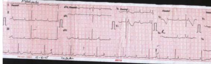
Figure: ECG shows sinus rhythm (P waves) at 120 bpm, independent of ventricular rhythm (QRS) at 45 bpm. No relationship between P waves and QRS complexes (AV dissociation). Narrow QRS escape rhythm (junctional).
💡 Examiner instruction (interactive): This is a case of Congenital Complete (Third-Degree) Heart Block – the most common cause of complete heart block in neonates, associated with maternal anti-Ro/SSA antibodies (SLE, Sjogren's). The candidate must recognize the ECG pattern (AV dissociation, independent P waves and QRS, atrial rate > ventricular rate), differentiate it from acquired causes (Lyme disease, post-surgical, myocarditis), and understand the indications for pacemaker (neonatal HR <55 bpm, wide QRS escape, structural CHD, ventricular dysfunction, symptoms). The candidate should also discuss the role of maternal corticosteroids (no proven benefit), the need for serial ECGs and echocardiograms, and the excellent prognosis with pacemaker placement.
🔍 Examiner Questions (interactive) – Click to reveal model answers
❓ Q1 (Examiner): “Describe the ECG findings in this infant. What is the diagnosis? How do you differentiate complete heart block from other types of AV block?”
✅ Candidate's answer:
• ECG findings:
- AV dissociation – no relationship between P waves and QRS complexes. Atrial rate (P waves) is faster than ventricular rate (QRS).
- Narrow QRS escape rhythm – suggests junctional (AV node) escape, which is more stable and has a better prognosis than a wide QRS (ventricular) escape.
- Regular P waves at a rate of 120 bpm (sinus rhythm).
- Regular QRS at a rate of 45 bpm – ventricular escape rhythm.
- No conduction from atria to ventricles.
• Diagnosis: Third-degree (complete) AV block.
• Differentiation from other AV blocks:
- First-degree: PR interval is prolonged (>0.18 sec) but all P waves are conducted.
- Second-degree Mobitz I: Progressive PR prolongation until a dropped QRS.
- Second-degree Mobitz II: Fixed PR interval with intermittent non-conducted P waves.
- Complete heart block: No P waves are conducted; AV dissociation is present.
• ECG findings:
- AV dissociation – no relationship between P waves and QRS complexes. Atrial rate (P waves) is faster than ventricular rate (QRS).
- Narrow QRS escape rhythm – suggests junctional (AV node) escape, which is more stable and has a better prognosis than a wide QRS (ventricular) escape.
- Regular P waves at a rate of 120 bpm (sinus rhythm).
- Regular QRS at a rate of 45 bpm – ventricular escape rhythm.
- No conduction from atria to ventricles.
• Diagnosis: Third-degree (complete) AV block.
• Differentiation from other AV blocks:
- First-degree: PR interval is prolonged (>0.18 sec) but all P waves are conducted.
- Second-degree Mobitz I: Progressive PR prolongation until a dropped QRS.
- Second-degree Mobitz II: Fixed PR interval with intermittent non-conducted P waves.
- Complete heart block: No P waves are conducted; AV dissociation is present.
❓ Q2 (Examiner): “What is the most common cause of congenital complete heart block in neonates? What is the role of maternal anti-Ro/SSA and anti-La/SSB antibodies?”
✅ Candidate's answer:
• Most common cause: Congenital Complete Heart Block (CHB) due to maternal anti-Ro/SSA (and anti-La/SSB) antibodies.
• Pathogenesis: Maternal antibodies cross the placenta and bind to fetal cardiac tissue (especially the AV node and His bundle), causing inflammation, fibrosis, and calcification of the conduction system. This usually occurs between 18-24 weeks of gestation.
• Maternal conditions: Systemic lupus erythematosus (SLE), Sjogren's syndrome, or asymptomatic antibody-positive mothers.
• Epidemiology: The risk of CHB in an infant of an anti-Ro-positive mother is approximately 1-2%. The recurrence risk in subsequent pregnancies is 15-20%.
• Other causes of congenital CHB: Structural congenital heart disease (L-transposition of great arteries, AVSD), maternal medications (rare).
• Prognosis: Without a pacemaker, 60-70% of neonates with CHB and a structurally normal heart survive, but the risk of sudden death is significant.
• Most common cause: Congenital Complete Heart Block (CHB) due to maternal anti-Ro/SSA (and anti-La/SSB) antibodies.
• Pathogenesis: Maternal antibodies cross the placenta and bind to fetal cardiac tissue (especially the AV node and His bundle), causing inflammation, fibrosis, and calcification of the conduction system. This usually occurs between 18-24 weeks of gestation.
• Maternal conditions: Systemic lupus erythematosus (SLE), Sjogren's syndrome, or asymptomatic antibody-positive mothers.
• Epidemiology: The risk of CHB in an infant of an anti-Ro-positive mother is approximately 1-2%. The recurrence risk in subsequent pregnancies is 15-20%.
• Other causes of congenital CHB: Structural congenital heart disease (L-transposition of great arteries, AVSD), maternal medications (rare).
• Prognosis: Without a pacemaker, 60-70% of neonates with CHB and a structurally normal heart survive, but the risk of sudden death is significant.
❓ Q3 (Examiner): “This infant's mother has SLE. Can fetal CHB be prevented? What is the role of maternal steroids, IVIG, or hydroxychloroquine?”
✅ Candidate's answer:
• Prevention of fetal CHB: Unfortunately, no proven interventions have been shown to reliably prevent fetal CHB in anti-Ro/La-positive pregnancies.
• Maternal steroids (dexamethasone): Some centers use dexamethasone (crosses the placenta) for treatment of fetal myocarditis or hydrops, but it has not been proven to prevent the development of CHB. It is associated with significant maternal and fetal side effects (IUGR, adrenal suppression).
- Indications for steroids: Fetal myocarditis, hydrops, or progression of first-degree to second-degree block.
• IVIG: Has been studied but not proven to prevent CHB.
• Hydroxychloroquine: Some studies suggest that hydroxychloroquine may reduce the incidence of CHB, but it is not established as a preventive therapy. However, it is recommended for maternal SLE management.
• Fetal monitoring: Weekly fetal echocardiograms from 16-26 weeks for anti-Ro-positive mothers. Early detection of first-degree block may allow for intervention (steroids) to prevent progression.
• Recurrence risk: 15-20% in subsequent pregnancies.
• Prevention of fetal CHB: Unfortunately, no proven interventions have been shown to reliably prevent fetal CHB in anti-Ro/La-positive pregnancies.
• Maternal steroids (dexamethasone): Some centers use dexamethasone (crosses the placenta) for treatment of fetal myocarditis or hydrops, but it has not been proven to prevent the development of CHB. It is associated with significant maternal and fetal side effects (IUGR, adrenal suppression).
- Indications for steroids: Fetal myocarditis, hydrops, or progression of first-degree to second-degree block.
• IVIG: Has been studied but not proven to prevent CHB.
• Hydroxychloroquine: Some studies suggest that hydroxychloroquine may reduce the incidence of CHB, but it is not established as a preventive therapy. However, it is recommended for maternal SLE management.
• Fetal monitoring: Weekly fetal echocardiograms from 16-26 weeks for anti-Ro-positive mothers. Early detection of first-degree block may allow for intervention (steroids) to prevent progression.
• Recurrence risk: 15-20% in subsequent pregnancies.
❓ Q4 (Examiner): “This infant has a heart rate of 45 bpm. What are the indications for pacemaker implantation in congenital CHB? When would you observe without a pacemaker?”
✅ Candidate's answer:
• Indications for pacemaker in congenital CHB (neonates):
1️⃣ Heart rate <55 bpm (especially in neonates).
2️⃣ Wide QRS escape rhythm (ventricular escape) – suggests infranodal block, worse prognosis.
3️⃣ Structural congenital heart disease (e.g., L-transposition, AVSD).
4️⃣ Ventricular dysfunction (impaired LVEF).
5️⃣ Symptoms – heart failure, poor feeding, tachypnea, syncope.
6️⃣ High-degree block with ventricular pauses >3 seconds.
7️⃣ Syncope – even a single episode.
• When to observe without a pacemaker:
- Heart rate >55 bpm, narrow QRS escape, normal ventricular function, no symptoms, no structural heart disease.
- In these cases, serial ECGs, Holter monitors, and echocardiograms are performed regularly.
- Observation is not without risk – sudden death can occur even in asymptomatic infants.
• Guidelines: Most pediatric electrophysiologists recommend pacemaker implantation in neonates with a narrow QRS and HR <55 bpm, and in all patients with a wide QRS escape rhythm, regardless of rate.
• Indications for pacemaker in congenital CHB (neonates):
1️⃣ Heart rate <55 bpm (especially in neonates).
2️⃣ Wide QRS escape rhythm (ventricular escape) – suggests infranodal block, worse prognosis.
3️⃣ Structural congenital heart disease (e.g., L-transposition, AVSD).
4️⃣ Ventricular dysfunction (impaired LVEF).
5️⃣ Symptoms – heart failure, poor feeding, tachypnea, syncope.
6️⃣ High-degree block with ventricular pauses >3 seconds.
7️⃣ Syncope – even a single episode.
• When to observe without a pacemaker:
- Heart rate >55 bpm, narrow QRS escape, normal ventricular function, no symptoms, no structural heart disease.
- In these cases, serial ECGs, Holter monitors, and echocardiograms are performed regularly.
- Observation is not without risk – sudden death can occur even in asymptomatic infants.
• Guidelines: Most pediatric electrophysiologists recommend pacemaker implantation in neonates with a narrow QRS and HR <55 bpm, and in all patients with a wide QRS escape rhythm, regardless of rate.
❓ Q5 (Examiner): “A 6-year-old after VSD closure has complete heart block on day 14. What is the management?”
✅ Candidate's answer:
• Diagnosis: Postoperative complete heart block (surgical injury to the conduction system during VSD closure).
• Management:
- Temporary epicardial pacing wires are typically placed during surgery.
- Observation for 10-14 days: If AV conduction returns, the patient may not need a permanent pacemaker.
- If CHB persists >10-14 days: Permanent pacemaker implantation is indicated.
- If the patient is symptomatic (syncope, hypotension) earlier, pacemaker is indicated sooner.
• Postoperative CHB and risk factors:
- VSD closure, AVSD repair, tetralogy of Fallot repair.
- Late-onset CHB: Can occur months or years after surgery – patients require long-term follow-up (ECG, Holter).
• Pacemaker type: Epicardial leads are preferred in small children; transvenous leads for older patients (>15 kg).
• Diagnosis: Postoperative complete heart block (surgical injury to the conduction system during VSD closure).
• Management:
- Temporary epicardial pacing wires are typically placed during surgery.
- Observation for 10-14 days: If AV conduction returns, the patient may not need a permanent pacemaker.
- If CHB persists >10-14 days: Permanent pacemaker implantation is indicated.
- If the patient is symptomatic (syncope, hypotension) earlier, pacemaker is indicated sooner.
• Postoperative CHB and risk factors:
- VSD closure, AVSD repair, tetralogy of Fallot repair.
- Late-onset CHB: Can occur months or years after surgery – patients require long-term follow-up (ECG, Holter).
• Pacemaker type: Epicardial leads are preferred in small children; transvenous leads for older patients (>15 kg).
❓ Q6 (Examiner): “A 12-year-old presents with erythema migrans rash, fever, and ECG showing complete heart block. What is the diagnosis and treatment?”
✅ Candidate's answer:
• Diagnosis: Lyme carditis (Borrelia burgdorferi) – can cause AV block (first-degree to complete).
• Clinical features: Erythema migrans, fever, arthralgia, headache. Heart block is a manifestation of disseminated Lyme disease.
• Management:
- IV Ceftriaxone: 50-75 mg/kg/day IV for 14-21 days (for carditis).
- Oral doxycycline: For localized Lyme disease without heart block (not for carditis).
- Temporary pacing: If symptomatic (syncope, hypotension) or high-degree block (Mobitz II, complete).
- Permanent pacemaker is rarely needed – the block typically resolves with antibiotics. Monitor closely; pacemaker only if block persists after treatment.
• Prognosis: Excellent with prompt treatment.
• Diagnosis: Lyme carditis (Borrelia burgdorferi) – can cause AV block (first-degree to complete).
• Clinical features: Erythema migrans, fever, arthralgia, headache. Heart block is a manifestation of disseminated Lyme disease.
• Management:
- IV Ceftriaxone: 50-75 mg/kg/day IV for 14-21 days (for carditis).
- Oral doxycycline: For localized Lyme disease without heart block (not for carditis).
- Temporary pacing: If symptomatic (syncope, hypotension) or high-degree block (Mobitz II, complete).
- Permanent pacemaker is rarely needed – the block typically resolves with antibiotics. Monitor closely; pacemaker only if block persists after treatment.
• Prognosis: Excellent with prompt treatment.
❓ Q7 (Examiner): “A child with acute myocarditis presents with complete heart block. How do you manage it?”
✅ Candidate's answer:
• Myocarditis can cause CHB due to inflammation of the conduction system.
• Management:
- Treat the underlying myocarditis – supportive care, heart failure management.
- Temporary pacing: If the patient is symptomatic (syncope, hypotension, heart failure) or has a very low escape rate.
- IVIG or corticosteroids – may be considered in autoimmune or giant cell myocarditis.
- Permanent pacemaker: Rarely needed – CHB often resolves with recovery of myocarditis. However, if the block persists >14 days or there is evidence of permanent conduction injury, a permanent pacemaker may be indicated.
- Follow-up: Serial ECGs, Holter monitors, and echocardiograms to monitor for recovery.
• Myocarditis can cause CHB due to inflammation of the conduction system.
• Management:
- Treat the underlying myocarditis – supportive care, heart failure management.
- Temporary pacing: If the patient is symptomatic (syncope, hypotension, heart failure) or has a very low escape rate.
- IVIG or corticosteroids – may be considered in autoimmune or giant cell myocarditis.
- Permanent pacemaker: Rarely needed – CHB often resolves with recovery of myocarditis. However, if the block persists >14 days or there is evidence of permanent conduction injury, a permanent pacemaker may be indicated.
- Follow-up: Serial ECGs, Holter monitors, and echocardiograms to monitor for recovery.
❓ Q8 (Examiner): “What genetic syndromes are associated with congenital heart block?”
✅ Candidate's answer:
• NKX2-5 mutations: Associated with secundum ASD and progressive AV block (first-degree → complete). Familial ASD with conduction disease.
• SCN5A mutations: Cause progressive cardiac conduction disease (Lenègre disease) – can present with first-degree, bundle branch block, and complete heart block. Associated with Brugada syndrome and LQTS3.
• Myotonic dystrophy type 1 (DM1): Progressive conduction disease (PR prolongation, QRS widening) and complete heart block.
• Emery-Dreifuss muscular dystrophy: Conduction defects, complete heart block, and cardiomyopathy.
• Kearns-Sayre syndrome (mitochondrial): Progressive conduction disease, complete heart block.
• L-transposition of the great arteries (ccTGA): Spontaneous CHB occurs at a rate of 2-5% per year due to abnormal location of the AV node.
• AVSD (Endocardial cushion defect): Associated with Down syndrome and CHB.
• NKX2-5 mutations: Associated with secundum ASD and progressive AV block (first-degree → complete). Familial ASD with conduction disease.
• SCN5A mutations: Cause progressive cardiac conduction disease (Lenègre disease) – can present with first-degree, bundle branch block, and complete heart block. Associated with Brugada syndrome and LQTS3.
• Myotonic dystrophy type 1 (DM1): Progressive conduction disease (PR prolongation, QRS widening) and complete heart block.
• Emery-Dreifuss muscular dystrophy: Conduction defects, complete heart block, and cardiomyopathy.
• Kearns-Sayre syndrome (mitochondrial): Progressive conduction disease, complete heart block.
• L-transposition of the great arteries (ccTGA): Spontaneous CHB occurs at a rate of 2-5% per year due to abnormal location of the AV node.
• AVSD (Endocardial cushion defect): Associated with Down syndrome and CHB.
❓ Q9 (Examiner): “This infant has congenital CHB. What is the recurrence risk for future pregnancies? How would you counsel the mother?”
✅ Candidate's answer:
• Recurrence risk: Approximately 15-20% for a subsequent pregnancy if the mother is positive for anti-Ro/SSA antibodies.
• Maternal counseling:
- The mother should be informed of the 15-20% recurrence risk.
- Fetal surveillance: Weekly fetal echocardiograms from 16-26 weeks gestation to monitor for the development of CHB.
- If fetal CHB is detected: Some centers use maternal dexamethasone (steroids) to treat fetal myocarditis and prevent progression, but this is controversial and not proven to reverse CHB.
- If first-degree block is detected: Early intervention with dexamethasone may prevent progression to complete heart block (some evidence).
- Delivery planning: Deliver in a tertiary care center with pediatric cardiology and electrophysiology support. The infant may require immediate pacemaker implantation after birth.
- Maternal medication: Hydroxychloroquine (for SLE) may reduce the risk of CHB and is recommended.
• Recurrence risk: Approximately 15-20% for a subsequent pregnancy if the mother is positive for anti-Ro/SSA antibodies.
• Maternal counseling:
- The mother should be informed of the 15-20% recurrence risk.
- Fetal surveillance: Weekly fetal echocardiograms from 16-26 weeks gestation to monitor for the development of CHB.
- If fetal CHB is detected: Some centers use maternal dexamethasone (steroids) to treat fetal myocarditis and prevent progression, but this is controversial and not proven to reverse CHB.
- If first-degree block is detected: Early intervention with dexamethasone may prevent progression to complete heart block (some evidence).
- Delivery planning: Deliver in a tertiary care center with pediatric cardiology and electrophysiology support. The infant may require immediate pacemaker implantation after birth.
- Maternal medication: Hydroxychloroquine (for SLE) may reduce the risk of CHB and is recommended.
❓ Q10 (Examiner): “What type of pacemaker is used in a neonate? What are the advantages of epicardial vs transvenous leads?”
✅ Candidate's answer:
• Epicardial pacing: Leads are placed on the epicardial surface of the heart (usually via a subxiphoid or thoracotomy approach).
- Indication: Preferred in infants and small children (< 15-20 kg) because of smaller vein size.
- Advantages: No need for transvenous access, less risk of venous thrombosis, no lead-related endocarditis.
- Disadvantages: Higher pacing thresholds, higher lead failure rate, more invasive placement, may need reoperation for lead replacement.
• Endocardial pacing (transvenous): Leads are placed via the subclavian vein into the right ventricle and/or right atrium.
- Indication: Preferred in older children/adolescents (> 15-20 kg).
- Advantages: Lower pacing thresholds, better lead durability, less invasive than epicardial.
- Disadvantages: Risk of venous thrombosis, pneumothorax, infection, lead displacement.
• Pacing modes: DDDR (dual-chamber rate-responsive) is preferred for active children to maintain AV synchrony.
• Leadless pacemaker: Micra (single-chamber) – available for larger children/adolescents.
• Epicardial pacing: Leads are placed on the epicardial surface of the heart (usually via a subxiphoid or thoracotomy approach).
- Indication: Preferred in infants and small children (< 15-20 kg) because of smaller vein size.
- Advantages: No need for transvenous access, less risk of venous thrombosis, no lead-related endocarditis.
- Disadvantages: Higher pacing thresholds, higher lead failure rate, more invasive placement, may need reoperation for lead replacement.
• Endocardial pacing (transvenous): Leads are placed via the subclavian vein into the right ventricle and/or right atrium.
- Indication: Preferred in older children/adolescents (> 15-20 kg).
- Advantages: Lower pacing thresholds, better lead durability, less invasive than epicardial.
- Disadvantages: Risk of venous thrombosis, pneumothorax, infection, lead displacement.
• Pacing modes: DDDR (dual-chamber rate-responsive) is preferred for active children to maintain AV synchrony.
• Leadless pacemaker: Micra (single-chamber) – available for larger children/adolescents.
❓ Q11 (Examiner): “What is the long-term prognosis for a neonate with congenital CHB?”
✅ Candidate's answer:
• Prognosis depends on:
- Presence of structural heart disease: Worse prognosis if associated with CHD (L-transposition, AVSD).
- Escape rhythm: Narrow QRS (junctional) escape has a better prognosis than wide QRS (ventricular) escape.
- Heart rate: HR <55 bpm at birth is associated with higher risk of heart failure and sudden death.
- Time to pacemaker: Early pacemaker implantation improves survival.
• With pacemaker therapy: Long-term survival is excellent (>90% at 10 years).
• Complications of pacemaker: Lead fracture, battery depletion, infection, superior vena cava syndrome (with transvenous leads).
• Need for ICD: Some patients with congenital CHB may develop ventricular arrhythmias (especially if associated with LV dysfunction).
• Transition to adult care: Lifelong follow-up with an electrophysiologist. Adult congenital heart disease (ACHD) specialists manage these patients.
• Prognosis depends on:
- Presence of structural heart disease: Worse prognosis if associated with CHD (L-transposition, AVSD).
- Escape rhythm: Narrow QRS (junctional) escape has a better prognosis than wide QRS (ventricular) escape.
- Heart rate: HR <55 bpm at birth is associated with higher risk of heart failure and sudden death.
- Time to pacemaker: Early pacemaker implantation improves survival.
• With pacemaker therapy: Long-term survival is excellent (>90% at 10 years).
• Complications of pacemaker: Lead fracture, battery depletion, infection, superior vena cava syndrome (with transvenous leads).
• Need for ICD: Some patients with congenital CHB may develop ventricular arrhythmias (especially if associated with LV dysfunction).
• Transition to adult care: Lifelong follow-up with an electrophysiologist. Adult congenital heart disease (ACHD) specialists manage these patients.
❓ Q12 (Examiner): “A 16-year-old with a pacemaker for congenital CHB wants to play basketball. What is the recommendation?”
✅ Candidate's answer:
• AHA/ACC guidelines for sports participation with a pacemaker:
- Non-contact sports (basketball, soccer, tennis) are generally allowed with precautions.
- Avoid contact sports (football, hockey, boxing, martial arts) – risk of damage to the device.
- Wear protective padding over the pacemaker pocket.
- Pre-participation evaluation: Ensure the pacemaker is functioning properly, and there is no underlying cardiomyopathy.
- Exercise test – assess heart rate response to exercise (if pacemaker is rate-responsive).
- Shared decision-making: Discuss risks and benefits. Most patients with pacemakers can lead active, normal lives.
• AHA/ACC guidelines for sports participation with a pacemaker:
- Non-contact sports (basketball, soccer, tennis) are generally allowed with precautions.
- Avoid contact sports (football, hockey, boxing, martial arts) – risk of damage to the device.
- Wear protective padding over the pacemaker pocket.
- Pre-participation evaluation: Ensure the pacemaker is functioning properly, and there is no underlying cardiomyopathy.
- Exercise test – assess heart rate response to exercise (if pacemaker is rate-responsive).
- Shared decision-making: Discuss risks and benefits. Most patients with pacemakers can lead active, normal lives.
❓ Q13 (Examiner): “Which drugs can cause complete heart block in children?”
✅ Candidate's answer:
• Common drugs that can cause AV block (dose-dependent):
- Digoxin: Toxicity can cause first-degree to complete heart block.
- Beta-blockers: Especially in high doses (propranolol, atenolol).
- Calcium channel blockers: Verapamil, diltiazem (can cause high-degree AV block).
- Antiarrhythmics: Class I (quinidine, procainamide), Class III (amiodarone, sotalol).
- Clonidine: Can cause bradycardia and AV block.
- Lithium: Can cause bradyarrhythmias.
- Neuromuscular blockers: Succinylcholine (rare).
- Magnesium: High doses can cause heart block.
• Management: Discontinue the offending drug. If symptomatic, temporary pacing may be needed. Atropine may be used for vagal-mediated block (if AV node level).
• Common drugs that can cause AV block (dose-dependent):
- Digoxin: Toxicity can cause first-degree to complete heart block.
- Beta-blockers: Especially in high doses (propranolol, atenolol).
- Calcium channel blockers: Verapamil, diltiazem (can cause high-degree AV block).
- Antiarrhythmics: Class I (quinidine, procainamide), Class III (amiodarone, sotalol).
- Clonidine: Can cause bradycardia and AV block.
- Lithium: Can cause bradyarrhythmias.
- Neuromuscular blockers: Succinylcholine (rare).
- Magnesium: High doses can cause heart block.
• Management: Discontinue the offending drug. If symptomatic, temporary pacing may be needed. Atropine may be used for vagal-mediated block (if AV node level).
❓ Q14 (Examiner): “The parents are very anxious about their infant's diagnosis. How will you counsel them?”
✅ Candidate's structured answer:
• “Your baby has a condition called congenital complete heart block – a rare condition where the electrical signals from the top of the heart do not reach the bottom chambers. This causes the heart to beat too slowly.”
• “The good news is that we have excellent treatments. Your baby will need a small device called a pacemaker – it sends electrical signals to make the heart beat at a normal rate. This is a very safe and effective procedure.”
• “The pacemaker will be placed either on the surface of the heart (epicardial) or through a vein (transvenous), depending on your baby's size. Most babies do very well after the pacemaker is placed.”
• “Your baby will need lifelong follow-up with a cardiologist to check the pacemaker, but most children with pacemakers lead active, normal lives.”
• “We will also monitor you closely in future pregnancies because there is a small risk (15-20%) of this happening again. We have a plan for fetal surveillance.”
• “You are not alone – we have a team of specialists who will support you and your baby every step of the way.”
• “Your baby has a condition called congenital complete heart block – a rare condition where the electrical signals from the top of the heart do not reach the bottom chambers. This causes the heart to beat too slowly.”
• “The good news is that we have excellent treatments. Your baby will need a small device called a pacemaker – it sends electrical signals to make the heart beat at a normal rate. This is a very safe and effective procedure.”
• “The pacemaker will be placed either on the surface of the heart (epicardial) or through a vein (transvenous), depending on your baby's size. Most babies do very well after the pacemaker is placed.”
• “Your baby will need lifelong follow-up with a cardiologist to check the pacemaker, but most children with pacemakers lead active, normal lives.”
• “We will also monitor you closely in future pregnancies because there is a small risk (15-20%) of this happening again. We have a plan for fetal surveillance.”
• “You are not alone – we have a team of specialists who will support you and your baby every step of the way.”
🗣️ Examiner's probing / high-yield points (Third-Degree Heart Block):
• "What is the most common cause of congenital CHB?" → Maternal anti-Ro/SSA antibodies.
• "What are the pacemaker indications in neonatal CHB?" → HR <55, wide QRS, structural CHD, symptoms, ventricular dysfunction.
• "What is the ECG finding in complete heart block?" → AV dissociation (P waves independent of QRS).
• "What is the recurrence risk for congenital CHB?" → 15-20%.
• "What is the treatment for Lyme carditis with CHB?" → IV ceftriaxone + temporary pacing if needed.
• "What genetic syndrome is associated with ASD and AV block?" → NKX2-5 mutation.
• "What type of pacemaker is used in neonates?" → Epicardial leads.
• "What is the prognosis for congenital CHB with a pacemaker?" → Excellent (>90% survival).
• "What is the most common cause of congenital CHB?" → Maternal anti-Ro/SSA antibodies.
• "What are the pacemaker indications in neonatal CHB?" → HR <55, wide QRS, structural CHD, symptoms, ventricular dysfunction.
• "What is the ECG finding in complete heart block?" → AV dissociation (P waves independent of QRS).
• "What is the recurrence risk for congenital CHB?" → 15-20%.
• "What is the treatment for Lyme carditis with CHB?" → IV ceftriaxone + temporary pacing if needed.
• "What genetic syndrome is associated with ASD and AV block?" → NKX2-5 mutation.
• "What type of pacemaker is used in neonates?" → Epicardial leads.
• "What is the prognosis for congenital CHB with a pacemaker?" → Excellent (>90% survival).
📘 Third-Degree (Complete) Heart Block – Core Revision for TOACS
⚡ Definition
Complete AV dissociation – no atrial impulses conducted to ventricles. Atrial rate > ventricular rate. Narrow QRS (junctional) escape vs wide QRS (ventricular) escape.
Complete AV dissociation – no atrial impulses conducted to ventricles. Atrial rate > ventricular rate. Narrow QRS (junctional) escape vs wide QRS (ventricular) escape.
🩺 Causes
Congenital: maternal anti-Ro/La antibodies (SLE, Sjogren's). Postoperative: VSD, AVSD, TOF repair. Acquired: Lyme disease, myocarditis, drugs (digoxin, beta-blockers), infiltrative.
Congenital: maternal anti-Ro/La antibodies (SLE, Sjogren's). Postoperative: VSD, AVSD, TOF repair. Acquired: Lyme disease, myocarditis, drugs (digoxin, beta-blockers), infiltrative.
🧬 Genetics
NKX2-5 (ASD + AV block), SCN5A (progressive conduction disease), myotonic dystrophy, Emery-Dreifuss, Kearns-Sayre.
NKX2-5 (ASD + AV block), SCN5A (progressive conduction disease), myotonic dystrophy, Emery-Dreifuss, Kearns-Sayre.
📊 ECG
AV dissociation, independent P waves and QRS, atrial rate > ventricular rate. Narrow QRS escape (better prognosis) vs wide QRS (worse).
AV dissociation, independent P waves and QRS, atrial rate > ventricular rate. Narrow QRS escape (better prognosis) vs wide QRS (worse).
💊 Management
Pacemaker if: HR <55 (neonate), wide QRS, structural CHD, symptoms, ventricular dysfunction. Lyme: IV ceftriaxone. Myocarditis: temporary pacing, treat underlying.
Pacemaker if: HR <55 (neonate), wide QRS, structural CHD, symptoms, ventricular dysfunction. Lyme: IV ceftriaxone. Myocarditis: temporary pacing, treat underlying.
📈 Pacemaker
Epicardial for infants (<15 kg), transvenous for older children. DDDR preferred. Lifelong follow-up.
Epicardial for infants (<15 kg), transvenous for older children. DDDR preferred. Lifelong follow-up.
⭐ High-yield pearls for TOACS (Third-Degree Heart Block):
• Most common cause (neonate): Maternal anti-Ro/SSA antibodies.
• ECG: AV dissociation, atrial rate > ventricular rate.
• Pacemaker if: HR <55, wide QRS, structural CHD, symptoms, LV dysfunction.
• Recurrence risk: 15-20%.
• Lyme carditis: IV ceftriaxone (14-21 days).
• NKX2-5: ASD + AV block.
• Epicardial leads for neonates.
• Most common cause (neonate): Maternal anti-Ro/SSA antibodies.
• ECG: AV dissociation, atrial rate > ventricular rate.
• Pacemaker if: HR <55, wide QRS, structural CHD, symptoms, LV dysfunction.
• Recurrence risk: 15-20%.
• Lyme carditis: IV ceftriaxone (14-21 days).
• NKX2-5: ASD + AV block.
• Epicardial leads for neonates.
🗣️ Candidate's role-play & examiner feedback
💬 To the candidate (role‑play): You will be asked the 14 questions from the Examiner Q&A tab. This station tests knowledge of Third-Degree (Complete) Heart Block – ECG interpretation (AV dissociation, atrial rate > ventricular rate), causes (congenital maternal anti-Ro/La, postoperative, Lyme, myocarditis), pacemaker indications (neonatal HR <55, wide QRS, structural CHD, symptoms), and management (temporary pacing, permanent pacemaker, treatment of underlying cause). Provide empathetic counseling to parents about prognosis, recurrence risk, and pacemaker therapy.
📝 Examiner Marking Grid (Third-Degree Heart Block – TOACS station):
- ✅ Interprets ECG: AV dissociation, independent P waves and QRS, atrial rate > ventricular rate
- ✅ Lists causes: congenital (anti-Ro/La), postoperative, Lyme, myocarditis, drugs
- ✅ States pacemaker indications: neonatal HR <55, wide QRS, structural CHD, symptoms, LV dysfunction
- ✅ Describes management of Lyme carditis: IV ceftriaxone ± temporary pacing
- ✅ Discusses postoperative CHB: temporary pacing, permanent if >10-14 days
- ✅ Explains genetics: NKX2-5 (ASD + AV block), SCN5A, myotonic dystrophy
- ✅ Describes pacemaker types: epicardial (neonates) vs transvenous (older)
- ✅ Discusses recurrence risk: 15-20% for congenital CHB
- ✅ Provides compassionate counseling and discusses prognosis
📚 Key references: Nelson Textbook of Pediatrics 22e (Chapter 484.7 – Atrioventricular Block), AHA/ACC/HRS Guidelines for Pacemaker, Pediatric EP Society recommendations.