FCPS Paediatrics TOACS · Interactive Station
🩺 Amniotic Band Syndrome (ABS) — Clinical recognition, limb deformity, management & counseling
📚 Paeds Online – paeds.online
⚕️ OBSERVED STATION · CPSP FORMAT · 8 MINUTES · SEPARATE TABS · AMNIOTIC BAND INCLUDED
📖 Problem-oriented Clinical Scenario + Image
👶🏻 Clinical Scenario (read aloud – 2 min):
A 2-hour-old term infant is brought to the newborn nursery for evaluation. The baby was born via spontaneous vaginal delivery at 39 weeks to a primigravida mother. Antenatal scans were reported as normal. On examination, the infant is hemodynamically stable. However, there is a deep circumferential constriction ring around the right ankle, with distal lymphoedema and dusky discoloration of the foot. The toes are intact but appear swollen. There is also pseudosyndactyly of two toes on the same foot—fused-looking soft tissue without bony union. No other anomalies are noted. The amniotic fluid was clear, and no other external abnormalities are present.
A clinical photograph of the constriction band is shown below.
Task: Describe the findings, propose the most likely diagnosis, discuss immediate management, and answer the examiner’s questions regarding etiology, associated anomalies, investigations, surgical timing, and long-term follow-up.
A 2-hour-old term infant is brought to the newborn nursery for evaluation. The baby was born via spontaneous vaginal delivery at 39 weeks to a primigravida mother. Antenatal scans were reported as normal. On examination, the infant is hemodynamically stable. However, there is a deep circumferential constriction ring around the right ankle, with distal lymphoedema and dusky discoloration of the foot. The toes are intact but appear swollen. There is also pseudosyndactyly of two toes on the same foot—fused-looking soft tissue without bony union. No other anomalies are noted. The amniotic fluid was clear, and no other external abnormalities are present.
A clinical photograph of the constriction band is shown below.
Task: Describe the findings, propose the most likely diagnosis, discuss immediate management, and answer the examiner’s questions regarding etiology, associated anomalies, investigations, surgical timing, and long-term follow-up.
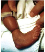
🔍 Figure: Amniotic band constriction ring around the right ankle. Note the deep groove, distal lymphoedema, and early changes of vascular compromise. Pseudosyndactyly of toes is also visible (soft tissue fusion). This is classical of Amniotic Band Sequence (ABS).
Source: PostImages – Band constricting the ankle.
Source: PostImages – Band constricting the ankle.
💡 Examiner instruction (interactive): The candidate will be asked to identify the condition, interpret the image, and then answer questions regarding pathophysiology, differential diagnosis, prenatal diagnosis, associated anomalies (including limb-body wall complex), surgical management (timing, techniques), rehabilitation, and counseling of parents.
🔍 Examiner Questions (interactive) – Click to reveal model answers
❓ Q1 (Examiner): “Describe the findings in the image and give the most likely diagnosis.”
✅ Candidate's structured answer:
• Findings: Deep circumferential constriction band around the right ankle, distal edema, dusky discoloration (suggesting venous/lymphatic compromise), and pseudosyndactyly of toes (soft tissue fusion without skeletal connection).
• Diagnosis: Amniotic Band Syndrome (ABS) / Amniotic Band Sequence (also known as Streeter dysplasia).
• Key point: This is a sporadic condition caused by early amnion rupture leading to entanglement of fetal parts by mesodermal bands.
• Findings: Deep circumferential constriction band around the right ankle, distal edema, dusky discoloration (suggesting venous/lymphatic compromise), and pseudosyndactyly of toes (soft tissue fusion without skeletal connection).
• Diagnosis: Amniotic Band Syndrome (ABS) / Amniotic Band Sequence (also known as Streeter dysplasia).
• Key point: This is a sporadic condition caused by early amnion rupture leading to entanglement of fetal parts by mesodermal bands.
❓ Q2 (Examiner): “What is the proposed pathogenesis of amniotic band syndrome?”
✅ Candidate's answer:
• Two main theories: (1) Exogenous theory – early amnion rupture (spontaneous) leads to formation of mesodential fibrous bands that entangle fetal parts, causing constriction rings, amputations, and clefts. (2) Endogenous theory (Streeter dysplasia) – primary germline defect in mesoderm formation; however, exogenous theory is more widely accepted.
• Constriction rings → impaired lymphatic/venous drainage → distal edema → possible ischemia, autoamputation.
• Associated with amnion rupture sequence and sometimes early oligohydramnios.
• Two main theories: (1) Exogenous theory – early amnion rupture (spontaneous) leads to formation of mesodential fibrous bands that entangle fetal parts, causing constriction rings, amputations, and clefts. (2) Endogenous theory (Streeter dysplasia) – primary germline defect in mesoderm formation; however, exogenous theory is more widely accepted.
• Constriction rings → impaired lymphatic/venous drainage → distal edema → possible ischemia, autoamputation.
• Associated with amnion rupture sequence and sometimes early oligohydramnios.
❓ Q3 (Examiner): “What are the associated congenital anomalies or syndromes linked to ABS?”
✅ Candidate's answer:
• Limb-body wall complex (severe form: thoracoabdominal schisis, exencephaly, limb defects).
• Craniofacial clefts (atypical facial clefts, encephalocele).
• Clubfoot, syndactyly, acrosyndactyly (pseudosyndactyly), terminal transverse limb defects.
• Constriction rings can affect digits, limbs, or even the umbilical cord (rare).
• Usually ABS is sporadic; not typically associated with chromosomal anomalies but rule out other anomalies via ultrasound.
• Limb-body wall complex (severe form: thoracoabdominal schisis, exencephaly, limb defects).
• Craniofacial clefts (atypical facial clefts, encephalocele).
• Clubfoot, syndactyly, acrosyndactyly (pseudosyndactyly), terminal transverse limb defects.
• Constriction rings can affect digits, limbs, or even the umbilical cord (rare).
• Usually ABS is sporadic; not typically associated with chromosomal anomalies but rule out other anomalies via ultrasound.
❓ Q4 (Examiner): “How can amniotic band syndrome be diagnosed prenatally?”
✅ Candidate's answer:
• High-resolution fetal ultrasound (2D/3D): visualization of thin, floating bands attached to fetal parts; constriction rings; asymmetric limb edema; amputations; associated clefts.
• Fetal MRI helpful for intracranial anomalies and bands not well seen on US.
• Key signs: “amniotic sheet” or bands, distal limb swelling, active fetal movement but with fixed deformity.
• Differential: limb body wall complex, early amnion rupture sequence.
• High-resolution fetal ultrasound (2D/3D): visualization of thin, floating bands attached to fetal parts; constriction rings; asymmetric limb edema; amputations; associated clefts.
• Fetal MRI helpful for intracranial anomalies and bands not well seen on US.
• Key signs: “amniotic sheet” or bands, distal limb swelling, active fetal movement but with fixed deformity.
• Differential: limb body wall complex, early amnion rupture sequence.
❓ Q5 (Examiner): “What are the differential diagnoses for a congenital constriction ring?”
✅ Candidate's answer:
• Amniotic Band Syndrome (most common).
• Streeter dysplasia (if considered separate entity).
• Congenital pseudoarthrosis (no circumferential band, more bony defect).
• Isolated congenital constriction bands (rare without other anomalies).
• Epidermolysis bullosa (pseudosyndactyly with blistering, but no amniotic bands).
• Vascular malformations causing limb edema but no ring.
• Amniotic Band Syndrome (most common).
• Streeter dysplasia (if considered separate entity).
• Congenital pseudoarthrosis (no circumferential band, more bony defect).
• Isolated congenital constriction bands (rare without other anomalies).
• Epidermolysis bullosa (pseudosyndactyly with blistering, but no amniotic bands).
• Vascular malformations causing limb edema but no ring.
❓ Q6 (Examiner): “What is your immediate postnatal evaluation for this infant?”
✅ Candidate's answer:
1️⃣ Complete physical examination – assess for other bands (digits, limbs, craniofacial, abdominal wall defects).
2️⃣ Assess limb perfusion – capillary refill distal to band, pulse oximetry, color, temperature, sensation (if possible).
3️⃣ Doppler ultrasound of extremity to evaluate arterial/venous flow.
4️⃣ Evaluate for other anomalies – echocardiogram, renal ultrasound if multiple anomalies present.
5️⃣ Baseline photographs and documentation, monitor for progression of ischemia.
1️⃣ Complete physical examination – assess for other bands (digits, limbs, craniofacial, abdominal wall defects).
2️⃣ Assess limb perfusion – capillary refill distal to band, pulse oximetry, color, temperature, sensation (if possible).
3️⃣ Doppler ultrasound of extremity to evaluate arterial/venous flow.
4️⃣ Evaluate for other anomalies – echocardiogram, renal ultrasound if multiple anomalies present.
5️⃣ Baseline photographs and documentation, monitor for progression of ischemia.
❓ Q7 (Examiner): “When is emergency surgical release of the band indicated in the newborn period?”
✅ Candidate's answer:
• Signs of vascular compromise – absent distal pulses, severe cyanosis, prolonged capillary refill (>5-7 sec), progressive ischemia.
• Compartment syndrome signs (pain, tense swelling, paresthesia – though difficult in neonate).
• Rapidly worsening edema with neurological deficit.
• In stable infant with good perfusion, surgery can be elective at 3-6 months.
• Emergency release via single-stage or two-stage Z-plasty to preserve limb.
• Signs of vascular compromise – absent distal pulses, severe cyanosis, prolonged capillary refill (>5-7 sec), progressive ischemia.
• Compartment syndrome signs (pain, tense swelling, paresthesia – though difficult in neonate).
• Rapidly worsening edema with neurological deficit.
• In stable infant with good perfusion, surgery can be elective at 3-6 months.
• Emergency release via single-stage or two-stage Z-plasty to preserve limb.
❓ Q8 (Examiner): “Describe the surgical management of a deep constriction band.”
✅ Candidate's answer:
• One-stage or two-stage Z-plasty – excision of the band and multiple interdigitating flaps to lengthen the circumference and release constriction.
• Microsurgical techniques to preserve neurovascular bundles.
• If severe ischemia at birth, emergency band release +/- fasciotomy.
• Two-stage procedure for deep bands (half the circumference released first, then 6-8 weeks later the other half) to avoid vascular compromise.
• Post-op splinting and hand/physiotherapy.
• One-stage or two-stage Z-plasty – excision of the band and multiple interdigitating flaps to lengthen the circumference and release constriction.
• Microsurgical techniques to preserve neurovascular bundles.
• If severe ischemia at birth, emergency band release +/- fasciotomy.
• Two-stage procedure for deep bands (half the circumference released first, then 6-8 weeks later the other half) to avoid vascular compromise.
• Post-op splinting and hand/physiotherapy.
❓ Q9 (Examiner): “How do you manage pseudosyndactyly (acrosyndactyly) associated with ABS?”
✅ Candidate's answer:
• Pseudosyndactyly is soft tissue fusion without bony continuity, often at the tips of digits.
• Surgical separation is indicated to improve function and prevent deformity.
• Usually performed after band release, around 6–12 months of age.
• Technique: separation of fused tissue, full-thickness skin grafting to create a web space.
• Requires hand surgeon/plastic surgeon experienced in congenital hand anomalies.
• Pseudosyndactyly is soft tissue fusion without bony continuity, often at the tips of digits.
• Surgical separation is indicated to improve function and prevent deformity.
• Usually performed after band release, around 6–12 months of age.
• Technique: separation of fused tissue, full-thickness skin grafting to create a web space.
• Requires hand surgeon/plastic surgeon experienced in congenital hand anomalies.
❓ Q10 (Examiner): “What non-surgical measures are important during the waiting period before surgery?”
✅ Candidate's answer:
• Monitor limb perfusion daily – color, capillary refill, temperature, limb circumference.
• Edema management – elevation of affected limb, gentle massage, compression garments (if tolerated).
• Prevent skin breakdown – keep the grooved area clean, emollients, avoid tight clothing.
• Parent education about signs of ischemia (darkening, coldness, pain with movement).
• Occupational/physiotherapy to maintain range of motion and prevent joint contractures.
• Monitor limb perfusion daily – color, capillary refill, temperature, limb circumference.
• Edema management – elevation of affected limb, gentle massage, compression garments (if tolerated).
• Prevent skin breakdown – keep the grooved area clean, emollients, avoid tight clothing.
• Parent education about signs of ischemia (darkening, coldness, pain with movement).
• Occupational/physiotherapy to maintain range of motion and prevent joint contractures.
❓ Q11 (Examiner): “What long-term follow-up is required for a child with ABS?”
✅ Candidate's answer:
• Plastic surgery / hand surgery clinic – serial evaluations for secondary procedures (recurrence of bands, scar contractures, web creep).
• Physical and occupational therapy – for functional use of limb, fine motor skills.
• Orthopedic assessment – for associated limb length discrepancy or joint deformities.
• Psychosocial support and family counseling regarding body image and developmental milestones.
• Genetic counseling – recurrence risk is extremely low (<1%) for sporadic ABS, but evaluate if familial clustering.
• Plastic surgery / hand surgery clinic – serial evaluations for secondary procedures (recurrence of bands, scar contractures, web creep).
• Physical and occupational therapy – for functional use of limb, fine motor skills.
• Orthopedic assessment – for associated limb length discrepancy or joint deformities.
• Psychosocial support and family counseling regarding body image and developmental milestones.
• Genetic counseling – recurrence risk is extremely low (<1%) for sporadic ABS, but evaluate if familial clustering.
❓ Q12 (Examiner): “What is the functional prognosis for an infant with a single deep ankle constriction band with mild distal edema?”
✅ Candidate's answer:
• Excellent prognosis if no vascular compromise and timely Z-plasty release.
• Most children achieve normal gait and activity after band release.
• If pseudosyndactyly is also corrected, near-normal toe function.
• However, residual cosmetic deformity (scar, slight circumferential indentation) may persist.
• Worse prognosis if associated with major anomalies (limb-body wall complex) or severe intrauterine amputation.
• Excellent prognosis if no vascular compromise and timely Z-plasty release.
• Most children achieve normal gait and activity after band release.
• If pseudosyndactyly is also corrected, near-normal toe function.
• However, residual cosmetic deformity (scar, slight circumferential indentation) may persist.
• Worse prognosis if associated with major anomalies (limb-body wall complex) or severe intrauterine amputation.
❓ Q13 (Examiner): “How will you counsel the parents regarding recurrence risk and future pregnancies?”
✅ Candidate's answer:
• Reassure that ABS is usually sporadic and not inherited; recurrence risk for future siblings is <1%.
• No specific prenatal prevention; however, advise detailed fetal anomaly scan at 18–22 weeks in subsequent pregnancies to look for amniotic bands.
• No association with maternal medications or lifestyle in most cases.
• Psychological support – emphasize that the child will lead a normal life after corrective procedures.
• Address guilt: “Nothing you did caused this; it is a random event during early gestation.”
• Reassure that ABS is usually sporadic and not inherited; recurrence risk for future siblings is <1%.
• No specific prenatal prevention; however, advise detailed fetal anomaly scan at 18–22 weeks in subsequent pregnancies to look for amniotic bands.
• No association with maternal medications or lifestyle in most cases.
• Psychological support – emphasize that the child will lead a normal life after corrective procedures.
• Address guilt: “Nothing you did caused this; it is a random event during early gestation.”
❓ Q14 (Examiner): “Is there a role for fetal intervention in amniotic band syndrome?”
✅ Candidate's answer:
• Fetoscopic laser release of constricting bands has been reported in selected cases with progressive limb ischemia or risk of autoamputation.
• Performed in specialized fetal therapy centers (similar to FETO for CDH).
• Indications: severe band causing edema/ischemia before viability, or progressive lymphedema with Doppler evidence of compromised flow.
• Risks: preterm premature rupture of membranes (PPROM), chorioamnionitis, preterm labor.
• Not universally recommended; most cases are managed postnatally.
• Fetoscopic laser release of constricting bands has been reported in selected cases with progressive limb ischemia or risk of autoamputation.
• Performed in specialized fetal therapy centers (similar to FETO for CDH).
• Indications: severe band causing edema/ischemia before viability, or progressive lymphedema with Doppler evidence of compromised flow.
• Risks: preterm premature rupture of membranes (PPROM), chorioamnionitis, preterm labor.
• Not universally recommended; most cases are managed postnatally.
❓ Q15 (Examiner): “What craniofacial anomalies can occur in ABS and how would you screen?”
✅ Candidate's answer:
• Atypical facial clefts (oblique facial clefts, Tessier clefts), naso-ocular clefts, cleft lip/palate.
• Encephalocele or microphthalmia.
• Screening: head ultrasound or MRI if neurological signs; clinical examination by craniofacial team; ophthalmologic exam for ocular anomalies.
• Audiology if craniofacial involvement (risk of hearing loss).
• In case of facial bands, refer to plastic surgery and speech therapy.
• Atypical facial clefts (oblique facial clefts, Tessier clefts), naso-ocular clefts, cleft lip/palate.
• Encephalocele or microphthalmia.
• Screening: head ultrasound or MRI if neurological signs; clinical examination by craniofacial team; ophthalmologic exam for ocular anomalies.
• Audiology if craniofacial involvement (risk of hearing loss).
• In case of facial bands, refer to plastic surgery and speech therapy.
🗣️ Examiner's probing / high-yield points:
• “What is the difference between pseudosyndactyly and true syndactyly?” → Pseudosyndactyly is soft tissue fusion at the tips, no bony or full web; true syndactyly has bony or full soft tissue union.
• “Can amniotic bands cause internal organ defects?” → Yes, in limb-body wall complex (abdominal/thoracic wall defects, heart, etc.).
• “What is the optimal timing for Z-plasty?” → Deep bands with lymphedema: often 3–6 months; emergency if ischemia.
• “What is the recurrence risk?” → Essentially nil; reassuring for parents.
• “Why is oligohydramnios not always present?” → ABS often occurs with normal fluid; early rupture may reseal.
• “What is the difference between pseudosyndactyly and true syndactyly?” → Pseudosyndactyly is soft tissue fusion at the tips, no bony or full web; true syndactyly has bony or full soft tissue union.
• “Can amniotic bands cause internal organ defects?” → Yes, in limb-body wall complex (abdominal/thoracic wall defects, heart, etc.).
• “What is the optimal timing for Z-plasty?” → Deep bands with lymphedema: often 3–6 months; emergency if ischemia.
• “What is the recurrence risk?” → Essentially nil; reassuring for parents.
• “Why is oligohydramnios not always present?” → ABS often occurs with normal fluid; early rupture may reseal.
📘 Amniotic Band Syndrome (ABS) – Core Revision
🔍 Definition
Amniotic Band Sequence: early amnion rupture → fibrous bands → entanglement → constriction rings, acrosyndactyly, amputations, clefts.
Amniotic Band Sequence: early amnion rupture → fibrous bands → entanglement → constriction rings, acrosyndactyly, amputations, clefts.
🧬 Etiology
Exogenous theory (amnion rupture) most accepted. Sporadic, not hereditary; recurrence <1%.
Exogenous theory (amnion rupture) most accepted. Sporadic, not hereditary; recurrence <1%.
📊 Associated anomalies
Limb-body wall complex, atypical facial clefts, encephalocele, clubfoot, transverse limb defects.
Limb-body wall complex, atypical facial clefts, encephalocele, clubfoot, transverse limb defects.
🩺 Prenatal Dx
USG: floating bands, constriction rings, distal edema, amputations. Fetal MRI adjunct.
USG: floating bands, constriction rings, distal edema, amputations. Fetal MRI adjunct.
⚕️ Management
Monitor perfusion. Surgery: Z-plasty release (one- or two-stage). Pseudosyndactyly release with grafting. Fetoscopic laser in severe selected cases.
Monitor perfusion. Surgery: Z-plasty release (one- or two-stage). Pseudosyndactyly release with grafting. Fetoscopic laser in severe selected cases.
📈 Follow-up
Hand/plastic surgery, PT/OT, assess for secondary deformities. Genetic counseling reassurance. Excellent prognosis for isolated bands.
Hand/plastic surgery, PT/OT, assess for secondary deformities. Genetic counseling reassurance. Excellent prognosis for isolated bands.
⭐ High-yield ABS pearls for TOACS:
• Clinical hallmark: Deep circumferential groove + distal edema + pseudosyndactyly.
• Do not confuse with: isolated congenital constriction ring (no bands elsewhere).
• Emergency release if signs of vascular compromise (absent pulses, blue/cold extremity).
• Acrosyndactyly = soft tissue fusion at tips; requires separation + skin graft.
• Counseling key: Sporadic, low recurrence, treatable surgically, good functional outcome.
• Clinical hallmark: Deep circumferential groove + distal edema + pseudosyndactyly.
• Do not confuse with: isolated congenital constriction ring (no bands elsewhere).
• Emergency release if signs of vascular compromise (absent pulses, blue/cold extremity).
• Acrosyndactyly = soft tissue fusion at tips; requires separation + skin graft.
• Counseling key: Sporadic, low recurrence, treatable surgically, good functional outcome.
🗣️ Candidate’s role-play & examiner feedback
💬 To the candidate (role‑play): You will be asked the 15 questions from the Examiner Q&A tab (including image interpretation, management, surgical details, counseling). Provide concise, evidence‑based answers. Examiner may ask for clarification on timing of surgery or differentials. Use structured points and be empathetic when counseling parents.
📝 Examiner Marking Grid (ABS – TOACS station):
- ✅ Correctly identifies the image: constriction band, pseudosyndactyly, amniotic band syndrome
- ✅ Explains pathogenesis (amnion rupture / exogenous theory)
- ✅ Lists associated anomalies (limb-body wall, facial clefts, etc.)
- ✅ Describes prenatal diagnosis (US + MRI)
- ✅ Recognizes emergency signs (vascular compromise) vs elective repair
- ✅ Knows surgical principles: Z-plasty (one- or two-stage), timing (3-6 months or urgent)
- ✅ Understands pseudosyndactyly management (separation + grafting)
- ✅ Provides appropriate long-term follow-up (therapy, surgical follow-up, psychosocial)
- ✅ Demonstrates empathetic counseling: sporadic, low recurrence, good prognosis
📚 Key references: Nelson Textbook of Pediatrics 22e, Ashcraft's Pediatric Surgery, CPSP guidelines on congenital anomalies, Journal of Pediatric Surgery (ABS fetoscopic release).