FCPS Paediatrics TOACS · Interactive Station
🩺 Congenital Hydrocephalus — Ventriculomegaly, macrocephaly, CSF diversion, VP shunt, neurodevelopmental outcome
📚 Paeds Online – paeds.online
⚕️ OBSERVED STATION · CPSP FORMAT · 8 MINUTES · SEPARATE TABS · CLINICAL PHOTO INCLUDED
📖 Problem-oriented Clinical Scenario + Photograph
👶🏻 Clinical Scenario (read aloud – 2 min):
A 2-day-old term male infant is transferred to the neonatal unit for evaluation of an enlarging head circumference. He was born at 39 weeks via normal vaginal delivery to a primigravida mother. No antenatal anomalies were noted. At birth, head circumference was 36 cm (90th percentile). Over 48 hours, the head circumference has increased to 38 cm, crossing percentiles. The anterior fontanel is full and tense, sutures are splayed, and the scalp veins are prominent. There is also a setting-sun sign (downward deviation of the eyes with sclera visible above the iris). The infant is irritable, has poor feeding, and intermittent emesis. The rest of the examination is normal.
A clinical photograph of the infant's head is shown below.
Task: Describe the findings, propose the most likely diagnosis, discuss the pathophysiology, classify the type of hydrocephalus, outline the diagnostic workup, and discuss management (medical vs surgical) including VP shunt complications.
A 2-day-old term male infant is transferred to the neonatal unit for evaluation of an enlarging head circumference. He was born at 39 weeks via normal vaginal delivery to a primigravida mother. No antenatal anomalies were noted. At birth, head circumference was 36 cm (90th percentile). Over 48 hours, the head circumference has increased to 38 cm, crossing percentiles. The anterior fontanel is full and tense, sutures are splayed, and the scalp veins are prominent. There is also a setting-sun sign (downward deviation of the eyes with sclera visible above the iris). The infant is irritable, has poor feeding, and intermittent emesis. The rest of the examination is normal.
A clinical photograph of the infant's head is shown below.
Task: Describe the findings, propose the most likely diagnosis, discuss the pathophysiology, classify the type of hydrocephalus, outline the diagnostic workup, and discuss management (medical vs surgical) including VP shunt complications.
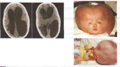
🔍 Figure: Newborn with congenital hydrocephalus – macrocephaly, prominent scalp veins, splayed cranial sutures (widened), and a bulging anterior fontanel. The setting-sun sign (downward deviation of eyes) is also a classic feature of increased intracranial pressure in infants.
💡 Examiner instruction (interactive): The candidate will be asked to identify congenital hydrocephalus, differentiate communicating from non-communicating types, discuss causes (aqueductal stenosis, Dandy-Walker, Chiari malformation), outline diagnostic imaging (head ultrasound, MRI), management (VP shunt, ETV), and discuss complications (shunt infection, obstruction, over-drainage) and neurodevelopmental outcomes.
🔍 Examiner Questions (interactive) – Click to reveal model answers
❓ Q1 (Examiner): “Describe the findings in the image. What is the most likely diagnosis? List the clinical signs of increased ICP in a newborn.”
✅ Candidate's structured answer:
• Findings: Macrocephaly (head circumference crossing percentiles), prominent scalp veins, splayed cranial sutures, tense anterior fontanel, setting-sun sign (inward-downward deviation of eyes).
• Diagnosis: Congenital hydrocephalus.
• Signs of increased ICP in newborn (with open sutures/fontanels):
- Rapid head growth, bulging fontanel, splayed sutures, sunset sign, irritability, vomiting, poor feeding, high-pitched cry, lethargy, apnea.
• Findings: Macrocephaly (head circumference crossing percentiles), prominent scalp veins, splayed cranial sutures, tense anterior fontanel, setting-sun sign (inward-downward deviation of eyes).
• Diagnosis: Congenital hydrocephalus.
• Signs of increased ICP in newborn (with open sutures/fontanels):
- Rapid head growth, bulging fontanel, splayed sutures, sunset sign, irritability, vomiting, poor feeding, high-pitched cry, lethargy, apnea.
❓ Q2 (Examiner): “Define hydrocephalus. What is the difference between communicating and non-communicating (obstructive) hydrocephalus?”
✅ Candidate's answer:
• Hydrocephalus: Abnormal accumulation of cerebrospinal fluid (CSF) within the ventricles (ventriculomegaly) due to impaired CSF flow, absorption, or rarely overproduction.
• Non-communicating (obstructive): Obstruction within the ventricular system (e.g., aqueductal stenosis, foramen of Monro atresia). CSF cannot reach the subarachnoid space. Most common congenital type.
• Communicating: No obstruction within ventricles; CSF flows out but absorption is impaired (e.g., post-hemorrhagic, post-meningitic, Chiari malformation, Dandy-Walker).
• Differentiation: By MRI/CT (ventricular dilation with/without obstruction) and sometimes by radionuclide cisternography.
• Hydrocephalus: Abnormal accumulation of cerebrospinal fluid (CSF) within the ventricles (ventriculomegaly) due to impaired CSF flow, absorption, or rarely overproduction.
• Non-communicating (obstructive): Obstruction within the ventricular system (e.g., aqueductal stenosis, foramen of Monro atresia). CSF cannot reach the subarachnoid space. Most common congenital type.
• Communicating: No obstruction within ventricles; CSF flows out but absorption is impaired (e.g., post-hemorrhagic, post-meningitic, Chiari malformation, Dandy-Walker).
• Differentiation: By MRI/CT (ventricular dilation with/without obstruction) and sometimes by radionuclide cisternography.
❓ Q3 (Examiner): “What are the common causes of congenital hydrocephalus?”
✅ Candidate's answer:
• Aqueductal stenosis (most common, 40%): Narrowing of cerebral aqueduct (Sylvius). Can be X-linked (L1CAM gene) or sporadic.
• Chiari II malformation: Associated with myelomeningocele → obstruction at fourth ventricle outlets.
• Dandy-Walker malformation: Cystic dilation of fourth ventricle, hypoplasia of cerebellar vermis, hydrocephalus.
• Post-hemorrhagic (IVH in preterm infants): Scarring → obstruction or impaired absorption.
• Post-infectious: Congenital infections (CMV, toxoplasmosis), neonatal meningitis.
• Neural tube defects (myelomeningocele) – associated Chiari II.
• Vein of Galen malformation (arteriovenous malformation).
• Chromosomal anomalies: Trisomy 13, 18, 21.
• Aqueductal stenosis (most common, 40%): Narrowing of cerebral aqueduct (Sylvius). Can be X-linked (L1CAM gene) or sporadic.
• Chiari II malformation: Associated with myelomeningocele → obstruction at fourth ventricle outlets.
• Dandy-Walker malformation: Cystic dilation of fourth ventricle, hypoplasia of cerebellar vermis, hydrocephalus.
• Post-hemorrhagic (IVH in preterm infants): Scarring → obstruction or impaired absorption.
• Post-infectious: Congenital infections (CMV, toxoplasmosis), neonatal meningitis.
• Neural tube defects (myelomeningocele) – associated Chiari II.
• Vein of Galen malformation (arteriovenous malformation).
• Chromosomal anomalies: Trisomy 13, 18, 21.
❓ Q4 (Examiner): “What imaging is indicated to diagnose hydrocephalus? What are the findings?”
✅ Candidate's answer:
• First-line: Cranial ultrasound (through anterior fontanel) – shows ventriculomegaly (enlarged lateral and third ventricles), periventricular lucency (edema), and can identify cause (IVH, aqueductal stenosis).
• MRI (or CT if MRI unavailable): Gold standard for anatomy, identifying the level of obstruction, associated anomalies (Chiari, Dandy-Walker), aqueductal stenosis, or mass lesions.
• Findings: Dilation of lateral ventricles (ballooning of frontal horns, "Mickey Mouse" ventricles), transependymal CSF flow (edema), third ventricle dilation, and sometimes periventricular leukomalacia.
• Measurement: Ventricular index (VI), frontal horn ratio, and assessment of progression.
• First-line: Cranial ultrasound (through anterior fontanel) – shows ventriculomegaly (enlarged lateral and third ventricles), periventricular lucency (edema), and can identify cause (IVH, aqueductal stenosis).
• MRI (or CT if MRI unavailable): Gold standard for anatomy, identifying the level of obstruction, associated anomalies (Chiari, Dandy-Walker), aqueductal stenosis, or mass lesions.
• Findings: Dilation of lateral ventricles (ballooning of frontal horns, "Mickey Mouse" ventricles), transependymal CSF flow (edema), third ventricle dilation, and sometimes periventricular leukomalacia.
• Measurement: Ventricular index (VI), frontal horn ratio, and assessment of progression.
❓ Q5 (Examiner): “What is the medical (non-surgical) management of hydrocephalus? Is it effective?”
✅ Candidate's answer:
• Medical therapy is rarely definitive and is used as a temporary measure or for mild, non-progressive hydrocephalus.
• Acetazolamide (carbonic anhydrase inhibitor) – reduces CSF production by up to 50%. Used in post-hemorrhagic hydrocephalus of prematurity as a temporizing measure. Side effects: metabolic acidosis, electrolyte imbalances.
• Furosemide (loop diuretic) – may be added.
• Serial lumbar punctures (LPs) – for post-hemorrhagic hydrocephalus in preterm infants to remove blood and protein, potentially preventing obstruction. May reduce need for shunt.
• Limitation: Medications not curative; surgery is definitive for progressive hydrocephalus with signs of increased ICP.
• Medical therapy is rarely definitive and is used as a temporary measure or for mild, non-progressive hydrocephalus.
• Acetazolamide (carbonic anhydrase inhibitor) – reduces CSF production by up to 50%. Used in post-hemorrhagic hydrocephalus of prematurity as a temporizing measure. Side effects: metabolic acidosis, electrolyte imbalances.
• Furosemide (loop diuretic) – may be added.
• Serial lumbar punctures (LPs) – for post-hemorrhagic hydrocephalus in preterm infants to remove blood and protein, potentially preventing obstruction. May reduce need for shunt.
• Limitation: Medications not curative; surgery is definitive for progressive hydrocephalus with signs of increased ICP.
❓ Q6 (Examiner): “What are the indications for VP shunt insertion in congenital hydrocephalus?”
✅ Candidate's answer:
• Absolute indications:
1️⃣ Progressive ventriculomegaly on serial imaging.
2️⃣ Signs of increased intracranial pressure (bulging fontanel, suture diastasis, sunset sign, vomiting, irritability, apnea).
3️⃣ Rapid head circumference growth crossing percentiles.
4️⃣ Transient or plateaued ventriculomegaly with neurological deterioration.
• Procedure: Insertion of a ventricular catheter into the lateral ventricle, connected to a unidirectional valve and peritoneal catheter (VP shunt). Other sites: ventriculoatrial (VA), ventriculopleural, or lumboperitoneal shunts.
• Goal: Divert CSF to the peritoneal cavity for absorption.
• Absolute indications:
1️⃣ Progressive ventriculomegaly on serial imaging.
2️⃣ Signs of increased intracranial pressure (bulging fontanel, suture diastasis, sunset sign, vomiting, irritability, apnea).
3️⃣ Rapid head circumference growth crossing percentiles.
4️⃣ Transient or plateaued ventriculomegaly with neurological deterioration.
• Procedure: Insertion of a ventricular catheter into the lateral ventricle, connected to a unidirectional valve and peritoneal catheter (VP shunt). Other sites: ventriculoatrial (VA), ventriculopleural, or lumboperitoneal shunts.
• Goal: Divert CSF to the peritoneal cavity for absorption.
❓ Q7 (Examiner): “What is endoscopic third ventriculostomy (ETV)? When is it preferred over a VP shunt?”
✅ Candidate's answer:
• Procedure: An endoscopic technique that creates a fenestration in the floor of the third ventricle, allowing CSF to flow directly into the subarachnoid space (bypassing the aqueduct). No shunt hardware.
• Best candidates: Non-communicating (obstructive) hydrocephalus, especially aqueductal stenosis. Also used in Dandy-Walker and Chiari (less effective).
• Advantages: No shunt-related complications (infection, obstruction, over-drainage), no lifelong foreign body.
• Disadvantages: Not effective in communicating hydrocephalus (poor CSF absorption), may fail over time (closure of stoma).
• Success rates: 60-80% for aqueductal stenosis; lower in infants <6 months of age.
• ETV failure requires VP shunt.
• Procedure: An endoscopic technique that creates a fenestration in the floor of the third ventricle, allowing CSF to flow directly into the subarachnoid space (bypassing the aqueduct). No shunt hardware.
• Best candidates: Non-communicating (obstructive) hydrocephalus, especially aqueductal stenosis. Also used in Dandy-Walker and Chiari (less effective).
• Advantages: No shunt-related complications (infection, obstruction, over-drainage), no lifelong foreign body.
• Disadvantages: Not effective in communicating hydrocephalus (poor CSF absorption), may fail over time (closure of stoma).
• Success rates: 60-80% for aqueductal stenosis; lower in infants <6 months of age.
• ETV failure requires VP shunt.
❓ Q8 (Examiner): “What are the common complications of VP shunts? How do they present?”
✅ Candidate's answer:
• Infection (5-10%): Usually due to Staphylococcus epidermidis or aureus. Presents with fever, irritability, erythema/swelling along shunt tract, abdominal pain, recurrent CSF pleocytosis. Requires shunt removal, external ventricular drain (EVD), and IV antibiotics.
• Obstruction (most common complication, 30-40% over 5 years): Blockage at proximal (ventricular) or distal (peritoneal) catheter. Presents with recurrent signs of increased ICP (vomiting, headache, bulging fontanel, sunset sign). Diagnosis: shunt series X-ray (to check discontinuity), CT/MRI (ventriculomegaly), shunt tap (to check patency).
• Over-drainage (slit ventricle syndrome): Chronic overdrainage → collapsed ventricles, intermittent headaches, vomiting, can lead to subdural hematoma.
• Under-drainage (shunt malfunction): Similar to obstruction.
• Abdominal complications: Perforation of bowel (peritonitis), pseudocyst formation (abdominal mass), ascites.
• Shunt migration or disconnection.
• Infection (5-10%): Usually due to Staphylococcus epidermidis or aureus. Presents with fever, irritability, erythema/swelling along shunt tract, abdominal pain, recurrent CSF pleocytosis. Requires shunt removal, external ventricular drain (EVD), and IV antibiotics.
• Obstruction (most common complication, 30-40% over 5 years): Blockage at proximal (ventricular) or distal (peritoneal) catheter. Presents with recurrent signs of increased ICP (vomiting, headache, bulging fontanel, sunset sign). Diagnosis: shunt series X-ray (to check discontinuity), CT/MRI (ventriculomegaly), shunt tap (to check patency).
• Over-drainage (slit ventricle syndrome): Chronic overdrainage → collapsed ventricles, intermittent headaches, vomiting, can lead to subdural hematoma.
• Under-drainage (shunt malfunction): Similar to obstruction.
• Abdominal complications: Perforation of bowel (peritonitis), pseudocyst formation (abdominal mass), ascites.
• Shunt migration or disconnection.
❓ Q9 (Examiner): “How would you recognize a VP shunt malfunction in an infant with previously well-controlled hydrocephalus?”
✅ Candidate's answer:
• Symptoms of increased ICP: Irritability, vomiting, lethargy, poor feeding, high-pitched cry, sunset sign, bulging fontanel, rapid head growth.
• Physical signs: Tense fontanel, splayed sutures, prominent scalp veins, setting-sun sign.
• Shunt site: Palpate the shunt reservoir (should refill quickly after pumping). Shunt tap by neurosurgeon.
• Imaging: CT/MRI showing increased ventricular size compared to baseline.
• Shunt series X-ray: Look for catheter disconnection or kinking.
• Any infant with a VP shunt and new onset vomiting/irritability should be presumed to have shunt malfunction until proven otherwise.
• Symptoms of increased ICP: Irritability, vomiting, lethargy, poor feeding, high-pitched cry, sunset sign, bulging fontanel, rapid head growth.
• Physical signs: Tense fontanel, splayed sutures, prominent scalp veins, setting-sun sign.
• Shunt site: Palpate the shunt reservoir (should refill quickly after pumping). Shunt tap by neurosurgeon.
• Imaging: CT/MRI showing increased ventricular size compared to baseline.
• Shunt series X-ray: Look for catheter disconnection or kinking.
• Any infant with a VP shunt and new onset vomiting/irritability should be presumed to have shunt malfunction until proven otherwise.
❓ Q10 (Examiner): “Can congenital hydrocephalus be diagnosed prenatally? What are the ultrasound findings?”
✅ Candidate's answer:
• Yes, diagnosed prenatally by ultrasound (second trimester).
• Findings: Ventriculomegaly (lateral ventricular atrium >10 mm at 18-22 weeks). Severe ventriculomegaly (>15 mm). May see dangling choroid plexus, enlarged third ventricle, and thinning of cerebral mantle.
• Associated anomalies: Neural tube defects (myelomeningocele), Dandy-Walker, aqueductal stenosis, Chiari malformation.
• Management: Detailed fetal MRI, fetal echocardiogram, amniocentesis for karyotype/microarray, CMV testing. Deliver at tertiary center with neurosurgery and NICU. Maternal-fetal counseling about prognosis (depends on etiology and severity).
• Fetal intervention: Rare; in utero ventriculoamniotic shunting not standard.
• Yes, diagnosed prenatally by ultrasound (second trimester).
• Findings: Ventriculomegaly (lateral ventricular atrium >10 mm at 18-22 weeks). Severe ventriculomegaly (>15 mm). May see dangling choroid plexus, enlarged third ventricle, and thinning of cerebral mantle.
• Associated anomalies: Neural tube defects (myelomeningocele), Dandy-Walker, aqueductal stenosis, Chiari malformation.
• Management: Detailed fetal MRI, fetal echocardiogram, amniocentesis for karyotype/microarray, CMV testing. Deliver at tertiary center with neurosurgery and NICU. Maternal-fetal counseling about prognosis (depends on etiology and severity).
• Fetal intervention: Rare; in utero ventriculoamniotic shunting not standard.
❓ Q11 (Examiner): “What is the neurodevelopmental prognosis for an infant with congenital hydrocephalus treated with VP shunt?”
✅ Candidate's answer:
• Outcome depends on etiology, timing of treatment, and associated anomalies.
• Isolated aqueductal stenosis treated early has better prognosis (~70-80% normal cognitive outcome).
• Hydrocephalus associated with myelomeningocele, Dandy-Walker, or prematurity has higher risk of neurodevelopmental impairment (NDI).
• Factors improving prognosis: Early diagnosis and shunting, absence of cortical thinning, no shunt infections, intact cerebral mantle.
• Sequelae:
- Cognitive deficits: 30-50% have learning disabilities or intellectual disability (mild to severe).
- Motor deficits: Spastic diplegia or quadriplegia (especially if associated with myelomeningocele or periventricular leukomalacia).
- Visual deficits: Strabismus, cortical visual impairment.
- Epilepsy (15-20%).
- Endocrine dysfunction: Precocious puberty (due to hypothalamic damage).
• Long-term multidisciplinary follow-up needed (neurosurgery, neurology, developmental pediatrics, ophthalmology).
• Outcome depends on etiology, timing of treatment, and associated anomalies.
• Isolated aqueductal stenosis treated early has better prognosis (~70-80% normal cognitive outcome).
• Hydrocephalus associated with myelomeningocele, Dandy-Walker, or prematurity has higher risk of neurodevelopmental impairment (NDI).
• Factors improving prognosis: Early diagnosis and shunting, absence of cortical thinning, no shunt infections, intact cerebral mantle.
• Sequelae:
- Cognitive deficits: 30-50% have learning disabilities or intellectual disability (mild to severe).
- Motor deficits: Spastic diplegia or quadriplegia (especially if associated with myelomeningocele or periventricular leukomalacia).
- Visual deficits: Strabismus, cortical visual impairment.
- Epilepsy (15-20%).
- Endocrine dysfunction: Precocious puberty (due to hypothalamic damage).
• Long-term multidisciplinary follow-up needed (neurosurgery, neurology, developmental pediatrics, ophthalmology).
❓ Q12 (Examiner): “What is slit ventricle syndrome? How does it present and how is it managed?”
✅ Candidate's answer:
• Definition: Chronic overdrainage of CSF causing the ventricles to collapse (become “slit-like”), leading to intermittent obstruction of the ventricular catheter, causing recurrent episodes of increased ICP.
• Presentation: Intermittent severe headaches, vomiting, lethargy, sometimes abducens nerve palsy (CN VI). CT shows slit-like ventricles (often normal-sized but symptomatic).
• Management:
- Antimigraine therapy (for headaches).
- Programmable (adjustable) shunt valve – set to higher pressure to prevent overdrainage.
- Antisiphon device – prevents siphoning when upright.
- Lumboperitoneal shunt or subtemporal decompression (rare).
- Avoid repeated shunt tapping (can worsen headaches).
• Can also cause subdural hematoma from tearing of bridging veins due to rapid overdrainage.
• Definition: Chronic overdrainage of CSF causing the ventricles to collapse (become “slit-like”), leading to intermittent obstruction of the ventricular catheter, causing recurrent episodes of increased ICP.
• Presentation: Intermittent severe headaches, vomiting, lethargy, sometimes abducens nerve palsy (CN VI). CT shows slit-like ventricles (often normal-sized but symptomatic).
• Management:
- Antimigraine therapy (for headaches).
- Programmable (adjustable) shunt valve – set to higher pressure to prevent overdrainage.
- Antisiphon device – prevents siphoning when upright.
- Lumboperitoneal shunt or subtemporal decompression (rare).
- Avoid repeated shunt tapping (can worsen headaches).
• Can also cause subdural hematoma from tearing of bridging veins due to rapid overdrainage.
❓ Q13 (Examiner): “How is VP shunt infection managed?”
✅ Candidate's answer:
• Step 1 – Remove the entire infected shunt (externalization). Place an external ventricular drain (EVD) for CSF diversion.
• Step 2 – Intravenous antibiotics (based on cultures, CSF gram stain). Common organisms: Coagulase-negative staphylococci, Staph aureus, gram-negative bacilli. Duration 10-14 days (some recommend 7 days after sterilization of CSF).
• Step 3 – Once CSF cultures are sterile (repeat LPs or EVD samples), insert a new VP shunt. Usually at a different site.
• Antibiotic-impregnated shunts reduce infection risk but do not eliminate it.
• Prognosis: Untreated infection can cause ventriculitis, meningitis, and poor neurodevelopmental outcomes.
• Step 1 – Remove the entire infected shunt (externalization). Place an external ventricular drain (EVD) for CSF diversion.
• Step 2 – Intravenous antibiotics (based on cultures, CSF gram stain). Common organisms: Coagulase-negative staphylococci, Staph aureus, gram-negative bacilli. Duration 10-14 days (some recommend 7 days after sterilization of CSF).
• Step 3 – Once CSF cultures are sterile (repeat LPs or EVD samples), insert a new VP shunt. Usually at a different site.
• Antibiotic-impregnated shunts reduce infection risk but do not eliminate it.
• Prognosis: Untreated infection can cause ventriculitis, meningitis, and poor neurodevelopmental outcomes.
❓ Q14 (Examiner): “How will you counsel the parents of this infant with congenital hydrocephalus regarding the need for VP shunt and long-term outcome?”
✅ Candidate's structured answer:
• “Your baby has hydrocephalus – a buildup of fluid in the brain’s ventricles causing pressure on the brain tissue and head enlargement. This is not anyone’s fault.”
• “The treatment is surgery to place a shunt – a thin tube that drains the extra fluid from the brain into the belly (peritoneum) where it is absorbed. This procedure is done by a pediatric neurosurgeon.”
• “The shunt is permanent, and it works very well in most cases. However, shunts can get blocked or infected (about 10% risk of infection). You will need to watch for signs of shunt malfunction – vomiting, irritability, bulging soft spot, sunset eyes – and come to the hospital immediately if these occur.”
• “The long-term outlook varies. Many children with isolated hydrocephalus have normal intelligence and lead normal lives. Some may have learning disabilities, motor problems, vision issues, or seizures. Early treatment improves outcomes.”
• “Your baby will need long-term follow-up with a team of specialists – neurosurgery, neurology, developmental pediatrics, ophthalmology, and physical therapy.”
• “We are here to support you through every step.”
• “Your baby has hydrocephalus – a buildup of fluid in the brain’s ventricles causing pressure on the brain tissue and head enlargement. This is not anyone’s fault.”
• “The treatment is surgery to place a shunt – a thin tube that drains the extra fluid from the brain into the belly (peritoneum) where it is absorbed. This procedure is done by a pediatric neurosurgeon.”
• “The shunt is permanent, and it works very well in most cases. However, shunts can get blocked or infected (about 10% risk of infection). You will need to watch for signs of shunt malfunction – vomiting, irritability, bulging soft spot, sunset eyes – and come to the hospital immediately if these occur.”
• “The long-term outlook varies. Many children with isolated hydrocephalus have normal intelligence and lead normal lives. Some may have learning disabilities, motor problems, vision issues, or seizures. Early treatment improves outcomes.”
• “Your baby will need long-term follow-up with a team of specialists – neurosurgery, neurology, developmental pediatrics, ophthalmology, and physical therapy.”
• “We are here to support you through every step.”
🗣️ Examiner's probing / high-yield points:
• "What is the most common cause of congenital hydrocephalus?" → Aqueductal stenosis (40%).
• "What is the difference between communicating and non-communicating hydrocephalus?" → Obstruction is within ventricles in non-communicating; absorption problem in communicating.
• "What is the setting-sun sign?" → Downward deviation of eyes with sclera visible above the iris – due to increased ICP in infants.
• "What is the first-line imaging?" → Cranial ultrasound (through fontanel).
• "What is the most common complication of VP shunt?" → Obstruction (30-40% over 5 years).
• "What is endoscopic third ventriculostomy (ETV)?" → Bypasses aqueductal stenosis; no shunt hardware.
• "What is the prognosis?" → Variable; depends on etiology. Isolated aqueductal stenosis has best outcome.
• "What is the most common cause of congenital hydrocephalus?" → Aqueductal stenosis (40%).
• "What is the difference between communicating and non-communicating hydrocephalus?" → Obstruction is within ventricles in non-communicating; absorption problem in communicating.
• "What is the setting-sun sign?" → Downward deviation of eyes with sclera visible above the iris – due to increased ICP in infants.
• "What is the first-line imaging?" → Cranial ultrasound (through fontanel).
• "What is the most common complication of VP shunt?" → Obstruction (30-40% over 5 years).
• "What is endoscopic third ventriculostomy (ETV)?" → Bypasses aqueductal stenosis; no shunt hardware.
• "What is the prognosis?" → Variable; depends on etiology. Isolated aqueductal stenosis has best outcome.
📘 Congenital Hydrocephalus – Core Revision for TOACS
🔍 Definition
Abnormal CSF accumulation within ventricles due to impaired flow, absorption, or (rarely) overproduction. Leads to ventriculomegaly and increased ICP.
Abnormal CSF accumulation within ventricles due to impaired flow, absorption, or (rarely) overproduction. Leads to ventriculomegaly and increased ICP.
📊 Classification
Non-communicating (obstructive – aqueductal stenosis, foramen atresia). Communicating (impaired absorption – post-hemorrhagic, post-meningitic).
Non-communicating (obstructive – aqueductal stenosis, foramen atresia). Communicating (impaired absorption – post-hemorrhagic, post-meningitic).
🩺 Clinical Signs
Macrocephaly (crossing percentiles), bulging fontanel, splayed sutures, sunset sign, irritability, vomiting, high-pitched cry.
Macrocephaly (crossing percentiles), bulging fontanel, splayed sutures, sunset sign, irritability, vomiting, high-pitched cry.
📷 Diagnosis
Cranial ultrasound (neonatal), MRI/CT. Ventricular index >97th percentile, transependymal edema.
Cranial ultrasound (neonatal), MRI/CT. Ventricular index >97th percentile, transependymal edema.
⚕️ Management
VP shunt (most common), ETV (for aqueductal stenosis). Temporizing: acetazolamide, serial LPs (in post-hemorrhagic).
VP shunt (most common), ETV (for aqueductal stenosis). Temporizing: acetazolamide, serial LPs (in post-hemorrhagic).
⚠️ Shunt Complications
Infection (5-10%, Staph epi), obstruction (30-40%, most common), over-drainage (slit ventricle syndrome), under-drainage, abdominal pseudocyst.
Infection (5-10%, Staph epi), obstruction (30-40%, most common), over-drainage (slit ventricle syndrome), under-drainage, abdominal pseudocyst.
📈 Prognosis
Depends on etiology. Isolated aqueductal stenosis → good. Associated with myelomeningocele, prematurity → higher risk of NDI (learning disability, motor deficits, epilepsy).
Depends on etiology. Isolated aqueductal stenosis → good. Associated with myelomeningocele, prematurity → higher risk of NDI (learning disability, motor deficits, epilepsy).
⭐ High-yield pearls for TOACS (Congenital Hydrocephalus):
• Most common cause: Aqueductal stenosis (X-linked or sporadic).
• First-line imaging: Cranial ultrasound (through fontanel).
• VP shunt complications: Obstruction (#1), infection (#2).
• Shunt infection organisms: Coagulase-negative staphylococci (S. epidermidis).
• ETV (Endoscopic third ventriculostomy): For aqueductal stenosis – no shunt.
• Setting-sun sign: Bilateral downward deviation of eyes – increased ICP.
• Rule out shunt malfunction: Any vomiting/irritability in a child with VP shunt.
• Most common cause: Aqueductal stenosis (X-linked or sporadic).
• First-line imaging: Cranial ultrasound (through fontanel).
• VP shunt complications: Obstruction (#1), infection (#2).
• Shunt infection organisms: Coagulase-negative staphylococci (S. epidermidis).
• ETV (Endoscopic third ventriculostomy): For aqueductal stenosis – no shunt.
• Setting-sun sign: Bilateral downward deviation of eyes – increased ICP.
• Rule out shunt malfunction: Any vomiting/irritability in a child with VP shunt.
🗣️ Candidate's role-play & examiner feedback
💬 To the candidate (role‑play): You will be asked the 14 questions from the Examiner Q&A tab (including clinical recognition, communicating vs non-communicating, imaging, VP shunt indications, complications, ETV, and parental counseling). Provide concise, evidence‑based answers. Examiner may ask about the setting-sun sign or shunt infection management. Use structured points and demonstrate empathy when counseling parents.
📝 Examiner Marking Grid (Congenital Hydrocephalus – TOACS station):
- ✅ Correctly identifies congenital hydrocephalus from image (macrocephaly, tense fontanel, sunset sign, splayed sutures)
- ✅ Differentiates communicating vs non-communicating hydrocephalus
- ✅ Lists common causes (aqueductal stenosis, Chiari II, Dandy-Walker, post-hemorrhagic)
- ✅ Recommends cranial ultrasound as first-line imaging
- ✅ Describes VP shunt insertion as definitive treatment for progressive hydrocephalus
- ✅ Discusses ETV (endoscopic third ventriculostomy) for aqueductal stenosis
- ✅ Lists VP shunt complications – obstruction (most common), infection (S. epidermidis), over-drainage
- ✅ States that shunt infection requires shunt removal + EVD + IV antibiotics
- ✅ Discusses variable prognosis (isolated aqueductal stenosis best, syndromic worse)
- ✅ Provides appropriate parental counseling (benign procedure, need for lifelong follow-up, signs of shunt malfunction)
📚 Key references: Nelson Textbook of Pediatrics 22e (Chapter 122.3 – Hydrocephalus), Volpe’s Neurology of the Newborn, CPSP guidelines on congenital CNS anomalies, Journal of Neurosurgery: Pediatrics.