FCPS Paediatrics TOACS · Interactive Station
🩺 Esophageal Atresia / Tracheoesophageal Fistula (EA/TEF) — Neonatal respiratory distress, feeding difficulty, surgical emergency
📚 Paeds Online – paeds.online
⚕️ OBSERVED STATION · CPSP FORMAT · 8 MINUTES · SEPARATE TABS · RADIOGRAPH INCLUDED
📖 Problem-oriented Clinical Scenario + Radiograph
👶🏻 Clinical Scenario (read aloud – 2 min):
A 2-hour-old term male infant is brought to the neonatal unit with excessive oral secretions and episodes of choking, coughing, and cyanosis immediately after birth. The baby was born at 39 weeks via normal vaginal delivery to a primigravida mother. Polyhydramnios was noted on antenatal ultrasound at 32 weeks. On examination: The infant is tachypneic (respiratory rate 70/min), with subcostal retractions and oxygen saturation 88% in room air. There is profuse, frothy, non-bilious salivation. An attempt to pass a nasogastric tube meets resistance at 10 cm from the gums, with the tube curling back in the mouth. The abdomen is scaphoid. Breath sounds are equal bilaterally with occasional crackles. A plain chest and abdominal radiograph is performed (shown below).
Task: Interpret the radiograph, propose the most likely diagnosis, classify the type of anomaly, and discuss immediate management.
A 2-hour-old term male infant is brought to the neonatal unit with excessive oral secretions and episodes of choking, coughing, and cyanosis immediately after birth. The baby was born at 39 weeks via normal vaginal delivery to a primigravida mother. Polyhydramnios was noted on antenatal ultrasound at 32 weeks. On examination: The infant is tachypneic (respiratory rate 70/min), with subcostal retractions and oxygen saturation 88% in room air. There is profuse, frothy, non-bilious salivation. An attempt to pass a nasogastric tube meets resistance at 10 cm from the gums, with the tube curling back in the mouth. The abdomen is scaphoid. Breath sounds are equal bilaterally with occasional crackles. A plain chest and abdominal radiograph is performed (shown below).
Task: Interpret the radiograph, propose the most likely diagnosis, classify the type of anomaly, and discuss immediate management.
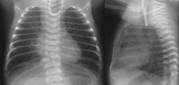
🔍 Figure: Chest and abdominal radiograph showing a coiled nasogastric tube in the proximal esophageal pouch (at T2-T3 level). The stomach and small bowel are distended with air, indicating a distal tracheoesophageal fistula (Gross type C). Absence of bowel gas would suggest pure esophageal atresia without fistula (type A).
Source: PostImages – Esophageal Atresia / TEF.
Source: PostImages – Esophageal Atresia / TEF.
💡 Examiner instruction (interactive): The candidate will be asked to interpret the X-ray, identify EA/TEF, classify the anomaly (Gross classification), discuss associated anomalies (VACTERL), immediate management (suction, positioning, IV fluids, antibiotics, surgical referral), and long-term complications (anastomotic leak, stricture, GERD, tracheomalacia).
🔍 Examiner Questions (interactive) – Click to reveal model answers
❓ Q1 (Examiner): “Describe the findings on this radiograph. What is the most likely diagnosis and the commonest type?”
✅ Candidate's structured answer:
• Radiograph findings: Nasogastric tube is coiled in the proximal esophagus (at T2-T3 level), indicating esophageal atresia. Bowel gas is present in the stomach and intestines (multiple air-filled loops), confirming a distal tracheoesophageal fistula (air travels from trachea through fistula into stomach).
• Diagnosis: Esophageal atresia with distal tracheoesophageal fistula (EA with distal TEF).
• Commonest type: Gross type C (EA with distal TEF) – accounts for ~85% of cases.
• Radiograph findings: Nasogastric tube is coiled in the proximal esophagus (at T2-T3 level), indicating esophageal atresia. Bowel gas is present in the stomach and intestines (multiple air-filled loops), confirming a distal tracheoesophageal fistula (air travels from trachea through fistula into stomach).
• Diagnosis: Esophageal atresia with distal tracheoesophageal fistula (EA with distal TEF).
• Commonest type: Gross type C (EA with distal TEF) – accounts for ~85% of cases.
❓ Q2 (Examiner): “Describe the Gross (Vogt) classification of esophageal atresia and tracheoesophageal fistula.”
✅ Candidate's answer:
• Type A (8%): Pure esophageal atresia (no fistula) – “long gap.” No bowel gas on X-ray.
• Type B (1%): EA with proximal TEF (fistula from proximal pouch to trachea) – rare.
• Type C (85%): EA with distal TEF (fistula from distal esophagus or trachea) – most common. Bowel gas present.
• Type D (1%): EA with both proximal and distal TEF (double fistula) – very rare.
• Type E (4%): Isolated TEF (H-type fistula) – no EA; presents later with choking, recurrent pneumonia.
• Type F: Congenital esophageal stenosis (very rare).
• Type A (8%): Pure esophageal atresia (no fistula) – “long gap.” No bowel gas on X-ray.
• Type B (1%): EA with proximal TEF (fistula from proximal pouch to trachea) – rare.
• Type C (85%): EA with distal TEF (fistula from distal esophagus or trachea) – most common. Bowel gas present.
• Type D (1%): EA with both proximal and distal TEF (double fistula) – very rare.
• Type E (4%): Isolated TEF (H-type fistula) – no EA; presents later with choking, recurrent pneumonia.
• Type F: Congenital esophageal stenosis (very rare).
❓ Q3 (Examiner): “What antenatal ultrasound findings suggest EA/TEF? What is the VACTERL association?”
✅ Candidate's answer:
• Antenatal findings: Polyhydramnios (inability to swallow amniotic fluid), small or absent fetal stomach bubble (in pure EA type A), occasionally dilated proximal esophageal pouch.
• VACTERL association (non-random association):
- V: Vertebral anomalies (hemivertebrae, scoliosis)
- A: Anal atresia / imperforate anus
- C: Cardiac defects (VSD, ASD, TOF, PDA)
- TE: Tracheo-Esophageal fistula / Esophageal atresia
- R: Renal anomalies (renal agenesis, hydronephrosis)
- L: Limb anomalies (radial ray defects, thumb hypoplasia, polydactyly)
• Key point: At least 3 of the above = VACTERL association. EA/TEF patients require screening for all these anomalies.
• Antenatal findings: Polyhydramnios (inability to swallow amniotic fluid), small or absent fetal stomach bubble (in pure EA type A), occasionally dilated proximal esophageal pouch.
• VACTERL association (non-random association):
- V: Vertebral anomalies (hemivertebrae, scoliosis)
- A: Anal atresia / imperforate anus
- C: Cardiac defects (VSD, ASD, TOF, PDA)
- TE: Tracheo-Esophageal fistula / Esophageal atresia
- R: Renal anomalies (renal agenesis, hydronephrosis)
- L: Limb anomalies (radial ray defects, thumb hypoplasia, polydactyly)
• Key point: At least 3 of the above = VACTERL association. EA/TEF patients require screening for all these anomalies.
❓ Q4 (Examiner): “What are the typical clinical features of EA/TEF in the newborn period?”
✅ Candidate's answer:
• Excessive oral secretions – frothy, non-bilious salivation (first sign).
• Choking, coughing, cyanosis with feeds – due to aspiration into trachea through fistula.
• Respiratory distress – tachypnea, retractions, crackles (aspiration pneumonia).
• Inability to pass nasogastric/orogastric tube – tube coils in upper pouch (confirmed by X-ray).
• Abdominal distention – occurs in type C (distal TEF) as air enters stomach via fistula.
• Scaphoid abdomen – in pure EA (type A) – no air in stomach.
• Excessive oral secretions – frothy, non-bilious salivation (first sign).
• Choking, coughing, cyanosis with feeds – due to aspiration into trachea through fistula.
• Respiratory distress – tachypnea, retractions, crackles (aspiration pneumonia).
• Inability to pass nasogastric/orogastric tube – tube coils in upper pouch (confirmed by X-ray).
• Abdominal distention – occurs in type C (distal TEF) as air enters stomach via fistula.
• Scaphoid abdomen – in pure EA (type A) – no air in stomach.
❓ Q5 (Examiner): “What is your immediate management in the delivery room and NICU before surgical repair?”
✅ Candidate's structured answer:
1️⃣ Keep NPO (nil per oral) – nothing by mouth.
2️⃣ Place a Replogle tube (double-lumen suction catheter) in the proximal esophageal pouch on continuous low suction to drain secretions and prevent aspiration.
3️⃣ Position head-up (30-45°) to reduce reflux of gastric contents through distal fistula.
4️⃣ Administer IV antibiotics (ampicillin + gentamicin) to cover aspiration pneumonia.
5️⃣ Secure IV access, maintain hydration with IVF.
6️⃣ Evaluate for associated anomalies – echocardiogram, renal ultrasound, spine X-ray, limb examination.
7️⃣ Stabilize respiratory status – avoid bag-mask ventilation (distends stomach through fistula, compromising ventilation). If ventilated, use low pressures and consider early gastrostomy to decompress stomach.
8️⃣ Refer to pediatric surgery for definitive repair.
1️⃣ Keep NPO (nil per oral) – nothing by mouth.
2️⃣ Place a Replogle tube (double-lumen suction catheter) in the proximal esophageal pouch on continuous low suction to drain secretions and prevent aspiration.
3️⃣ Position head-up (30-45°) to reduce reflux of gastric contents through distal fistula.
4️⃣ Administer IV antibiotics (ampicillin + gentamicin) to cover aspiration pneumonia.
5️⃣ Secure IV access, maintain hydration with IVF.
6️⃣ Evaluate for associated anomalies – echocardiogram, renal ultrasound, spine X-ray, limb examination.
7️⃣ Stabilize respiratory status – avoid bag-mask ventilation (distends stomach through fistula, compromising ventilation). If ventilated, use low pressures and consider early gastrostomy to decompress stomach.
8️⃣ Refer to pediatric surgery for definitive repair.
❓ Q6 (Examiner): “When is surgical repair performed? Describe the operative approach.”
✅ Candidate's answer:
• Timing: After stabilization, typically within 24-48 hours of birth. Delayed repair if extreme prematurity, severe pneumonia, or major cardiac anomalies.
• Approach: Right extrapleural thoracotomy (4th-5th intercostal space) or thoracoscopic repair in select centers.
• Procedure: Division of the tracheoesophageal fistula, closure of tracheal defect, and primary end-to-end esophageal anastomosis (if gap length permits).
• Long-gap EA: May require delayed primary repair, esophageal substitution (gastric pull-up, colonic interposition), or Foker technique (traction-induced growth).
• Gastrostomy tube placed in selected cases for decompression or feeding.
• Timing: After stabilization, typically within 24-48 hours of birth. Delayed repair if extreme prematurity, severe pneumonia, or major cardiac anomalies.
• Approach: Right extrapleural thoracotomy (4th-5th intercostal space) or thoracoscopic repair in select centers.
• Procedure: Division of the tracheoesophageal fistula, closure of tracheal defect, and primary end-to-end esophageal anastomosis (if gap length permits).
• Long-gap EA: May require delayed primary repair, esophageal substitution (gastric pull-up, colonic interposition), or Foker technique (traction-induced growth).
• Gastrostomy tube placed in selected cases for decompression or feeding.
❓ Q7 (Examiner): “What are the common early and late complications of EA/TEF repair?”
✅ Candidate's answer:
• Early complications:
- Anastomotic leak (5-10%) – managed conservatively with chest tube drainage, nil per oral, parenteral nutrition.
- Recurrent TEF (3-5%) – presents with recurrent pneumonia, choking; requires repeat repair.
- Pneumothorax, chylothorax (thoracic duct injury).
- Vocal cord paralysis (recurrent laryngeal nerve injury) – hoarse cry, aspiration risk.
• Late complications:
- Anastomotic stricture (20-40%) – presents with dysphagia, food impaction; treated with esophageal dilation.
- Gastroesophageal reflux disease (GERD) – very common (>50%); may require fundoplication.
- Tracheomalacia (compression by innominate artery) – barking cough, cyanotic spells (“dying spells”); may require aortopexy.
- Esophageal dysmotility – lifelong difficulty swallowing large boluses.
- Recurrent aspiration pneumonia, reactive airways disease.
• Early complications:
- Anastomotic leak (5-10%) – managed conservatively with chest tube drainage, nil per oral, parenteral nutrition.
- Recurrent TEF (3-5%) – presents with recurrent pneumonia, choking; requires repeat repair.
- Pneumothorax, chylothorax (thoracic duct injury).
- Vocal cord paralysis (recurrent laryngeal nerve injury) – hoarse cry, aspiration risk.
• Late complications:
- Anastomotic stricture (20-40%) – presents with dysphagia, food impaction; treated with esophageal dilation.
- Gastroesophageal reflux disease (GERD) – very common (>50%); may require fundoplication.
- Tracheomalacia (compression by innominate artery) – barking cough, cyanotic spells (“dying spells”); may require aortopexy.
- Esophageal dysmotility – lifelong difficulty swallowing large boluses.
- Recurrent aspiration pneumonia, reactive airways disease.
❓ Q8 (Examiner): “How do you diagnose and manage an anastomotic stricture?”
✅ Candidate's answer:
• Presentation: Dysphagia (difficulty swallowing, feeding refusal), choking, recurrent vomiting, failure to thrive. Typically appears 4-8 weeks post-repair.
• Diagnosis: Upper GI contrast study (barium swallow) shows narrowing at anastomosis; esophagoscopy confirms.
• Management: Esophageal dilation (balloon or bougie). Most require 1-3 dilations. Refractory strictures may need local steroid injection or resection.
• Prevention: Some centers use prophylactic antireflux medications and early dilations.
• Presentation: Dysphagia (difficulty swallowing, feeding refusal), choking, recurrent vomiting, failure to thrive. Typically appears 4-8 weeks post-repair.
• Diagnosis: Upper GI contrast study (barium swallow) shows narrowing at anastomosis; esophagoscopy confirms.
• Management: Esophageal dilation (balloon or bougie). Most require 1-3 dilations. Refractory strictures may need local steroid injection or resection.
• Prevention: Some centers use prophylactic antireflux medications and early dilations.
❓ Q9 (Examiner): “Why is GERD common after EA/TEF repair and how is it managed?”
✅ Candidate's answer:
• Why common? Impaired esophageal motility, short intra-abdominal esophagus, abnormal angle of His, and associated vagal nerve injury during surgery.
• Medical management: Positioning (head-up, prone after feeds), thickened feeds, proton pump inhibitors (PPIs) or H2 blockers (ranitidine).
• Surgical management (fundoplication): Indicated for severe GERD with failure to thrive, recurrent aspiration, stricture refractory to dilation, or recurrent TEF.
• Note: Fundoplication may worsen dysphagia in some due to underlying dysmotility.
• Why common? Impaired esophageal motility, short intra-abdominal esophagus, abnormal angle of His, and associated vagal nerve injury during surgery.
• Medical management: Positioning (head-up, prone after feeds), thickened feeds, proton pump inhibitors (PPIs) or H2 blockers (ranitidine).
• Surgical management (fundoplication): Indicated for severe GERD with failure to thrive, recurrent aspiration, stricture refractory to dilation, or recurrent TEF.
• Note: Fundoplication may worsen dysphagia in some due to underlying dysmotility.
❓ Q10 (Examiner): “What is tracheomalacia in EA/TEF and when is aortopexy indicated?”
✅ Candidate's answer:
• Tracheomalacia: Weakness of tracheal cartilage where the TEF was located, causing dynamic airway collapse during expiration or crying.
• Clinical features: “Barking” or “brassy” cough, expiratory stridor, recurrent cyanotic spells (“dying spells”) – sudden apnea and cyanosis due to complete tracheal collapse.
• Aortopexy: Surgical suspension of the innominate artery (and aorta) to the sternum to lift the anterior trachea open. Indicated for severe, life-threatening episodes not responding to conservative management.
• Other measures: Continuous positive airway pressure (CPAP) may stent open the trachea; expectant management – most improve by 2-3 years as tracheal cartilage matures.
• Tracheomalacia: Weakness of tracheal cartilage where the TEF was located, causing dynamic airway collapse during expiration or crying.
• Clinical features: “Barking” or “brassy” cough, expiratory stridor, recurrent cyanotic spells (“dying spells”) – sudden apnea and cyanosis due to complete tracheal collapse.
• Aortopexy: Surgical suspension of the innominate artery (and aorta) to the sternum to lift the anterior trachea open. Indicated for severe, life-threatening episodes not responding to conservative management.
• Other measures: Continuous positive airway pressure (CPAP) may stent open the trachea; expectant management – most improve by 2-3 years as tracheal cartilage matures.
❓ Q11 (Examiner): “What feeding and nutritional support is required after EA/TEF repair?”
✅ Candidate's answer:
• Postoperative: NPO for 5-7 days; parenteral nutrition. Obtain esophagram on day 5-7 to check for leak; if normal, start enteral feeds.
• Oral feeding difficulties: Common due to esophageal dysmotility. Start with continuous nasogastric feeds, transition to bolus, then oral.
• Gastrostomy tube may be placed in long-gap EA or severe GERD.
• Long-term: Monitor growth; small frequent meals, semi-solid foods often better tolerated. Speech and feeding therapy for oral aversion.
• Failure to thrive: Look for stricture, GERD, recurrent TEF. Caloric supplementation (modular formula) may be needed.
• Postoperative: NPO for 5-7 days; parenteral nutrition. Obtain esophagram on day 5-7 to check for leak; if normal, start enteral feeds.
• Oral feeding difficulties: Common due to esophageal dysmotility. Start with continuous nasogastric feeds, transition to bolus, then oral.
• Gastrostomy tube may be placed in long-gap EA or severe GERD.
• Long-term: Monitor growth; small frequent meals, semi-solid foods often better tolerated. Speech and feeding therapy for oral aversion.
• Failure to thrive: Look for stricture, GERD, recurrent TEF. Caloric supplementation (modular formula) may be needed.
❓ Q12 (Examiner): “What long-term respiratory problems occur in EA/TEF survivors?”
✅ Candidate's answer:
• Tracheomalacia (improves with age but may persist into childhood).
• Recurrent aspiration pneumonia – due to GERD, TEF recurrence, or vocal cord dysfunction.
• Reactive airway disease / asthma-like symptoms – wheezing, chronic cough (high incidence).
• Bronchiectasis – from repeated aspiration/infection.
• Follow-up: Pulmonology referral, pulmonary function testing (when older), immunization (including influenza, RSV prophylaxis in high-risk).
• Tracheomalacia (improves with age but may persist into childhood).
• Recurrent aspiration pneumonia – due to GERD, TEF recurrence, or vocal cord dysfunction.
• Reactive airway disease / asthma-like symptoms – wheezing, chronic cough (high incidence).
• Bronchiectasis – from repeated aspiration/infection.
• Follow-up: Pulmonology referral, pulmonary function testing (when older), immunization (including influenza, RSV prophylaxis in high-risk).
❓ Q13 (Examiner): “What investigations are needed to screen for associated anomalies in a newborn with EA/TEF?”
✅ Candidate's answer:
• Echocardiogram – to rule out congenital heart disease (VSD, TOF, PDA, ASD).
• Renal ultrasound – for renal agenesis, hydronephrosis, cystic kidneys.
• Spine X-ray or ultrasound – vertebral anomalies (hemivertebrae, scoliosis).
• Abdominal ultrasound – for duodenal atresia, malrotation, biliary anomalies.
• Limb examination and X-rays – radial ray defects, thumb hypoplasia, polydactyly.
• Imperforate anus – careful perineal exam; abdominal X-ray to rule out distal intestinal obstruction.
• Chromosomal microarray – if syndromic features (Feingold syndrome, CHARGE, 22q11 deletion).
• Echocardiogram – to rule out congenital heart disease (VSD, TOF, PDA, ASD).
• Renal ultrasound – for renal agenesis, hydronephrosis, cystic kidneys.
• Spine X-ray or ultrasound – vertebral anomalies (hemivertebrae, scoliosis).
• Abdominal ultrasound – for duodenal atresia, malrotation, biliary anomalies.
• Limb examination and X-rays – radial ray defects, thumb hypoplasia, polydactyly.
• Imperforate anus – careful perineal exam; abdominal X-ray to rule out distal intestinal obstruction.
• Chromosomal microarray – if syndromic features (Feingold syndrome, CHARGE, 22q11 deletion).
❓ Q14 (Examiner): “What is the prognosis for an infant with isolated EA/TEF (type C)? How will you counsel the parents?”
✅ Candidate's answer:
• Survival: Excellent (>95%) for isolated EA/TEF with modern neonatal and surgical care. Survival decreases significantly if associated with complex cardiac anomalies or extreme prematurity.
• Functional outcome: Most children lead normal lives but require long-term surveillance for GERD, strictures, and respiratory issues.
• Feeding and growth: Many have some degree of dysphagia (especially solids), but most achieve normal nutrition with small, frequent meals.
• Recurrence risk: Low (<1%) for isolated EA/TEF; higher if part of a genetic syndrome. No specific folic acid prevention (unlike neural tube defects).
• Counseling points: Reassure parents that outcome is good but they should be prepared for possible dilations, GERD medication, and feeding challenges. Emphasize importance of long-term multidisciplinary follow-up (pediatric surgery, gastroenterology, pulmonology, nutrition, speech therapy).
• Survival: Excellent (>95%) for isolated EA/TEF with modern neonatal and surgical care. Survival decreases significantly if associated with complex cardiac anomalies or extreme prematurity.
• Functional outcome: Most children lead normal lives but require long-term surveillance for GERD, strictures, and respiratory issues.
• Feeding and growth: Many have some degree of dysphagia (especially solids), but most achieve normal nutrition with small, frequent meals.
• Recurrence risk: Low (<1%) for isolated EA/TEF; higher if part of a genetic syndrome. No specific folic acid prevention (unlike neural tube defects).
• Counseling points: Reassure parents that outcome is good but they should be prepared for possible dilations, GERD medication, and feeding challenges. Emphasize importance of long-term multidisciplinary follow-up (pediatric surgery, gastroenterology, pulmonology, nutrition, speech therapy).
🗣️ Examiner's probing / high-yield points:
• "What is the ‘VACTERL’ association and why is it important?" → Vertebral, Anal, Cardiac, TE fistula, Renal, Limb. Every EA/TEF infant must be screened for these.
• "Why is bowel gas on X-ray important in EA/TEF?" → Presence of gas indicates distal TEF (type C). No gas suggests pure atresia (type A) or proximal TEF.
• "What is a Replogle tube and why is it used?" → Double-lumen suction catheter placed in proximal pouch for continuous drainage of saliva to prevent aspiration.
• "How do you differentiate EA/TEF from isolated esophageal stenosis?" → Stenosis presents later (>6 months) with dysphagia and food impaction; X-ray shows no coiled tube.
• "What is the most common cause of late death in EA/TEF?" → Aspiration pneumonia or severe GERD complications.
• "What is the ‘VACTERL’ association and why is it important?" → Vertebral, Anal, Cardiac, TE fistula, Renal, Limb. Every EA/TEF infant must be screened for these.
• "Why is bowel gas on X-ray important in EA/TEF?" → Presence of gas indicates distal TEF (type C). No gas suggests pure atresia (type A) or proximal TEF.
• "What is a Replogle tube and why is it used?" → Double-lumen suction catheter placed in proximal pouch for continuous drainage of saliva to prevent aspiration.
• "How do you differentiate EA/TEF from isolated esophageal stenosis?" → Stenosis presents later (>6 months) with dysphagia and food impaction; X-ray shows no coiled tube.
• "What is the most common cause of late death in EA/TEF?" → Aspiration pneumonia or severe GERD complications.
📘 Esophageal Atresia / Tracheoesophageal Fistula – Core Revision
🔍 Definition
Congenital discontinuity of esophagus with or without fistulous connection to trachea. Incidence: 1 in 3,000-4,000 live births.
Congenital discontinuity of esophagus with or without fistulous connection to trachea. Incidence: 1 in 3,000-4,000 live births.
📊 Gross Classification
Type C (EA with distal TEF) = 85%. Type A (pure EA) = 8%. Type E (H-type fistula) = 4%.
Type C (EA with distal TEF) = 85%. Type A (pure EA) = 8%. Type E (H-type fistula) = 4%.
🧬 Associations
VACTERL (Vertebral, Anal, Cardiac, TE fistula, Renal, Limb). Polyhydramnios antenatally.
VACTERL (Vertebral, Anal, Cardiac, TE fistula, Renal, Limb). Polyhydramnios antenatally.
🩺 Clinical Dx
Excessive salivation, choking with feeds, NG tube coils on X-ray. Bowel gas = distal TEF.
Excessive salivation, choking with feeds, NG tube coils on X-ray. Bowel gas = distal TEF.
⚕️ Pre-op Management
Replogle tube suction, head-up position, NPO, IV antibiotics, avoid bag-mask ventilation.
Replogle tube suction, head-up position, NPO, IV antibiotics, avoid bag-mask ventilation.
📈 Surgical Repair
Right thoracotomy (or thoracoscopy), division of fistula, primary anastomosis. Gap length determines complexity.
Right thoracotomy (or thoracoscopy), division of fistula, primary anastomosis. Gap length determines complexity.
⚠️ Complications
Anastomotic leak, stricture (20-40%), GERD (>50%), recurrent TEF (3-5%), tracheomalacia, vocal cord paralysis.
Anastomotic leak, stricture (20-40%), GERD (>50%), recurrent TEF (3-5%), tracheomalacia, vocal cord paralysis.
🔁 Long-term
GERD management, serial dilations, aortopexy for severe tracheomalacia, pulmonary follow-up, growth monitoring.
GERD management, serial dilations, aortopexy for severe tracheomalacia, pulmonary follow-up, growth monitoring.
⭐ High-yield pearls for TOACS (EA/TEF):
• X-ray hallmark: Coiled nasogastric tube in upper pouch + bowel gas (distal TEF) = type C.
• Never feed the infant. Place Replogle tube on continuous suction immediately.
• VACTERL screening is mandatory – echocardiogram, renal ultrasound, spine imaging, perineal exam.
• Post-op stricture is common → presents with dysphagia; treat with esophageal dilation.
• Tracheomalacia spells (cyanotic episodes) may require aortopexy.
• Prognosis excellent for isolated EA/TEF; long-term GERD and respiratory issues need surveillance.
• X-ray hallmark: Coiled nasogastric tube in upper pouch + bowel gas (distal TEF) = type C.
• Never feed the infant. Place Replogle tube on continuous suction immediately.
• VACTERL screening is mandatory – echocardiogram, renal ultrasound, spine imaging, perineal exam.
• Post-op stricture is common → presents with dysphagia; treat with esophageal dilation.
• Tracheomalacia spells (cyanotic episodes) may require aortopexy.
• Prognosis excellent for isolated EA/TEF; long-term GERD and respiratory issues need surveillance.
🗣️ Candidate’s role-play & examiner feedback
💬 To the candidate (role‑play): You will be asked the 14 questions from the Examiner Q&A tab (including X-ray interpretation, Gross classification, VACTERL association, immediate management, surgical principles, complications, and long-term follow-up). Provide concise, evidence‑based answers. Examiner may ask for clarification on Replogle tube use or tracheomalacia management. Use structured points and demonstrate systematic thinking.
📝 Examiner Marking Grid (EA/TEF – TOACS station):
- ✅ Correctly interprets X-ray (coiled NG tube + bowel gas → type C EA/TEF)
- ✅ Lists Gross classification (A to E) with approximate frequencies
- ✅ Describes VACTERL association and necessary screening
- ✅ Recognizes clinical presentation (polyhydramnios, frothy secretions, choking with feeds)
- ✅ Outlines immediate pre-op management (Replogle tube, NPO, head-up, antibiotics, avoid bag-mask)
- ✅ Knows surgical approach (right thoracotomy, fistula division, anastomosis)
- ✅ Lists early complications (leak, recurrent TEF, pneumothorax, vocal cord palsy)
- ✅ Discusses late complications (stricture, GERD, tracheomalacia, dysmotility)
- ✅ Describes management of stricture (dilation) and tracheomalacia (aortopexy if severe)
- ✅ Provides appropriate long-term follow-up and prognosis counseling
📚 Key references: Nelson Textbook of Pediatrics 22e, Ashcraft's Pediatric Surgery, Gross classification of EA/TEF, CPSP guidelines on neonatal surgical emergencies, VACTERL association.