FCPS Paediatrics TOACS · Interactive Station
🩺 Umbilical Granuloma — Clinical recognition, differential diagnosis, management & parental counseling
📚 Paeds Online – paeds.online
⚕️ OBSERVED STATION · CPSP FORMAT · 8 MINUTES · SEPARATE TABS · CLINICAL IMAGE INCLUDED
📖 Problem-oriented Clinical Scenario + Photograph
👶🏻 Clinical Scenario (read aloud – 2 min):
A 3-week-old term infant is brought to the pediatric outpatient clinic by the mother. The baby was born at 39 weeks via normal vaginal delivery, birth weight 3.2 kg. The umbilical cord separated at day 10 without any bleeding or discharge. For the past 10 days, the mother has noticed a moist, pinkish-red lump at the umbilicus. It produces a small amount of clear, sticky discharge. There is no foul odor, no fever, and the baby is feeding well, active, and afebrile. On examination, the infant is well-looking. The umbilicus shows a soft, friable, granulomatous lesion ~6 mm in diameter at the base of the umbilical stump. The surrounding skin is normal with no erythema, warmth, or induration. No other abnormalities are noted.
A clinical photograph of the umbilical lesion is shown below.
Task: Describe the findings, propose the most likely diagnosis, discuss the differential diagnosis, and answer the examiner’s questions regarding management, complications, and parental advice.
A 3-week-old term infant is brought to the pediatric outpatient clinic by the mother. The baby was born at 39 weeks via normal vaginal delivery, birth weight 3.2 kg. The umbilical cord separated at day 10 without any bleeding or discharge. For the past 10 days, the mother has noticed a moist, pinkish-red lump at the umbilicus. It produces a small amount of clear, sticky discharge. There is no foul odor, no fever, and the baby is feeding well, active, and afebrile. On examination, the infant is well-looking. The umbilicus shows a soft, friable, granulomatous lesion ~6 mm in diameter at the base of the umbilical stump. The surrounding skin is normal with no erythema, warmth, or induration. No other abnormalities are noted.
A clinical photograph of the umbilical lesion is shown below.
Task: Describe the findings, propose the most likely diagnosis, discuss the differential diagnosis, and answer the examiner’s questions regarding management, complications, and parental advice.
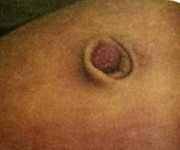
🔍 Figure: Well-defined, pink, moist, granulomatous lesion at the umbilicus. Typical appearance of an umbilical granuloma after cord separation. No surrounding cellulitis or discharge.
Source: PostImages – Umbilical Granuloma.
Source: PostImages – Umbilical Granuloma.
💡 Examiner instruction (interactive): The candidate will be asked to identify the condition, describe the image, discuss etiology, differential diagnoses (patent urachus, omphalomesenteric duct remnant, omphalitis), outline treatment options (silver nitrate cauterization, saline wet-to-dry dressings, surgical excision), and provide parental counseling regarding complications and when to refer.
🔍 Examiner Questions (interactive) – Click to reveal model answers
❓ Q1 (Examiner): “Describe the lesion shown in the image. What is the most likely diagnosis?”
✅ Candidate's structured answer:
• Findings: Solitary, pinkish-red, moist, friable, soft nodule at the umbilical base. Size ~5-10 mm. No surrounding erythema or purulent discharge.
• Diagnosis: Umbilical granuloma – a common benign overgrowth of granulation tissue at the umbilicus after separation of the cord.
• Key point: Occurs due to delayed or incomplete epithelialization of the umbilical stump.
• Findings: Solitary, pinkish-red, moist, friable, soft nodule at the umbilical base. Size ~5-10 mm. No surrounding erythema or purulent discharge.
• Diagnosis: Umbilical granuloma – a common benign overgrowth of granulation tissue at the umbilicus after separation of the cord.
• Key point: Occurs due to delayed or incomplete epithelialization of the umbilical stump.
❓ Q2 (Examiner): “What is the pathophysiology of umbilical granuloma?”
✅ Candidate's answer:
• After cord separation (usually 1-2 weeks), the base should heal by epithelialization.
• Failure of complete epithelialization leads to formation of exuberant granulation tissue (rich in capillaries and fibroblasts).
• Chronic mild inflammation and moisture promote persistence.
• It is not a true congenital anomaly; rather a postnatal healing disorder.
• After cord separation (usually 1-2 weeks), the base should heal by epithelialization.
• Failure of complete epithelialization leads to formation of exuberant granulation tissue (rich in capillaries and fibroblasts).
• Chronic mild inflammation and moisture promote persistence.
• It is not a true congenital anomaly; rather a postnatal healing disorder.
❓ Q3 (Examiner): “What are the important differential diagnoses of an umbilical granuloma?”
✅ Candidate's answer:
1️⃣ Patent urachus – clear urine discharge, connection to bladder, ultrasound or sinogram shows tract.
2️⃣ Omphalomesenteric (vitelline) duct remnant – persistent discharge of intestinal fluid, may contain meconium or stool; can prolapse as a polyp (bright red, mucosal).
3️⃣ Omphalitis (umbilical infection) – erythema, induration, purulent discharge, fever, systemic signs.
4️⃣ Umbilical polyp (remnant of omphalomesenteric duct) – often more firm, dark red, may be covered by intestinal mucosa.
5️⃣ Pyogenic granuloma (rare at umbilicus).
• Key investigation to rule out patent urachus or vitelline duct: ultrasound of umbilicus and bladder, or voiding cystourethrogram (VCUG) / fistulogram.
1️⃣ Patent urachus – clear urine discharge, connection to bladder, ultrasound or sinogram shows tract.
2️⃣ Omphalomesenteric (vitelline) duct remnant – persistent discharge of intestinal fluid, may contain meconium or stool; can prolapse as a polyp (bright red, mucosal).
3️⃣ Omphalitis (umbilical infection) – erythema, induration, purulent discharge, fever, systemic signs.
4️⃣ Umbilical polyp (remnant of omphalomesenteric duct) – often more firm, dark red, may be covered by intestinal mucosa.
5️⃣ Pyogenic granuloma (rare at umbilicus).
• Key investigation to rule out patent urachus or vitelline duct: ultrasound of umbilicus and bladder, or voiding cystourethrogram (VCUG) / fistulogram.
❓ Q4 (Examiner): “What clinical features would raise suspicion of a patent urachus or omphalomesenteric duct remnant instead of simple granuloma?”
✅ Candidate's answer:
• Persistent or intermittent clear (urine) discharge → patent urachus.
• Fecal or bilious discharge → omphalomesenteric duct remnant.
• Recurrent granuloma after adequate silver nitrate cauterization (3–4 treatments).
• Large (>1 cm) or polypoid lesion that looks like intestinal mucosa.
• Associated abdominal wall defects or malformations.
• Presence of urinary symptoms or umbilical hernia with discharge.
• Persistent or intermittent clear (urine) discharge → patent urachus.
• Fecal or bilious discharge → omphalomesenteric duct remnant.
• Recurrent granuloma after adequate silver nitrate cauterization (3–4 treatments).
• Large (>1 cm) or polypoid lesion that looks like intestinal mucosa.
• Associated abdominal wall defects or malformations.
• Presence of urinary symptoms or umbilical hernia with discharge.
❓ Q5 (Examiner): “What is the first-line conservative treatment for umbilical granuloma before considering chemical cautery?”
✅ Candidate's answer:
• Dry cord care + topical 70% isopropyl alcohol or saline – cleaning 2-3 times daily.
• Application of common salt (table salt / sodium chloride) – a pinch of salt covered with dry gauze for 30 minutes twice daily leads to desiccation and regression of granulation tissue. Effective and inexpensive.
• Observation – some small granulomas may resolve spontaneously by 4-6 weeks.
• If no improvement after 1-2 weeks of salt therapy, proceed to silver nitrate cauterization.
• Dry cord care + topical 70% isopropyl alcohol or saline – cleaning 2-3 times daily.
• Application of common salt (table salt / sodium chloride) – a pinch of salt covered with dry gauze for 30 minutes twice daily leads to desiccation and regression of granulation tissue. Effective and inexpensive.
• Observation – some small granulomas may resolve spontaneously by 4-6 weeks.
• If no improvement after 1-2 weeks of salt therapy, proceed to silver nitrate cauterization.
❓ Q6 (Examiner): “Describe the correct technique for silver nitrate cauterization of umbilical granuloma. Include precautions.”
✅ Candidate's answer:
• Consent and explain procedure to parents.
• Protect surrounding skin with petroleum jelly (Vaseline) or zinc oxide barrier to prevent chemical burns.
• Use silver nitrate applicator stick (moisten tip with sterile water or saline to activate).
• Gently roll/touch the applicator to the granuloma only, avoiding normal skin.
• Lesion turns white/grey immediately → chemical cauterization.
• Apply for 10–20 seconds; repeat in 1-2 weeks if needed (max 3-4 sessions).
• Precautions: avoid deep penetration, do not use on >1 cm lesions, avoid if infection suspected. Warn parents about transient discoloration.
• Consent and explain procedure to parents.
• Protect surrounding skin with petroleum jelly (Vaseline) or zinc oxide barrier to prevent chemical burns.
• Use silver nitrate applicator stick (moisten tip with sterile water or saline to activate).
• Gently roll/touch the applicator to the granuloma only, avoiding normal skin.
• Lesion turns white/grey immediately → chemical cauterization.
• Apply for 10–20 seconds; repeat in 1-2 weeks if needed (max 3-4 sessions).
• Precautions: avoid deep penetration, do not use on >1 cm lesions, avoid if infection suspected. Warn parents about transient discoloration.
❓ Q7 (Examiner): “What are the contraindications to silver nitrate cauterization of umbilical granuloma?”
✅ Candidate's answer:
• Signs of omphalitis (cellulitis, purulent discharge, fever).
• Suspicion of patent urachus or vitelline duct remnant (risk of intraperitoneal spill, chemical peritonitis).
• Large granuloma (>1 cm) or polypoid morphology – refer for surgical evaluation.
• Known allergy to silver (rare).
• Uncooperative infant (risk of skin burn) – but can be managed with immobilization.
• Signs of omphalitis (cellulitis, purulent discharge, fever).
• Suspicion of patent urachus or vitelline duct remnant (risk of intraperitoneal spill, chemical peritonitis).
• Large granuloma (>1 cm) or polypoid morphology – refer for surgical evaluation.
• Known allergy to silver (rare).
• Uncooperative infant (risk of skin burn) – but can be managed with immobilization.
❓ Q8 (Examiner): “What do you do if the granuloma recurs after 3 sessions of silver nitrate cauterization?”
✅ Candidate's answer:
• Suspect an underlying remnant (patent urachus or omphalomesenteric duct).
• Perform umbilical ultrasound to assess for tract or cystic structure.
• If necessary, sinogram or fistulogram (contrast study).
• Refer to pediatric surgery for possible surgical excision under general anesthesia.
• Excision and closure of the base – simple surgical removal with histopathology to confirm diagnosis.
• Suspect an underlying remnant (patent urachus or omphalomesenteric duct).
• Perform umbilical ultrasound to assess for tract or cystic structure.
• If necessary, sinogram or fistulogram (contrast study).
• Refer to pediatric surgery for possible surgical excision under general anesthesia.
• Excision and closure of the base – simple surgical removal with histopathology to confirm diagnosis.
❓ Q9 (Examiner): “What are the potential complications of an untreated or improperly treated umbilical granuloma?”
✅ Candidate's answer:
• Persistent moist discharge → local skin maceration and irritation.
• Secondary bacterial infection → omphalitis, cellulitis, possible necrotizing fasciitis (rare but serious).
• Bleeding from friable granulation tissue (minor).
• Parental anxiety and repeated clinic visits.
• If underlying urachal remnant is missed, risk of urachal cyst, recurrent urinary tract infections, or urachal adenocarcinoma later in life (very rare).
• Persistent moist discharge → local skin maceration and irritation.
• Secondary bacterial infection → omphalitis, cellulitis, possible necrotizing fasciitis (rare but serious).
• Bleeding from friable granulation tissue (minor).
• Parental anxiety and repeated clinic visits.
• If underlying urachal remnant is missed, risk of urachal cyst, recurrent urinary tract infections, or urachal adenocarcinoma later in life (very rare).
❓ Q10 (Examiner): “When is surgical excision indicated as primary treatment?”
✅ Candidate's answer:
• Large granuloma (>1 cm) that is unlikely to respond to cautery.
• Polypoid lesion suspicious for omphalomesenteric duct remnant.
• Persistent discharge despite adequate chemical cauterization (2-3 sessions).
• Confirmed patent urachus or vitelline duct on imaging.
• Procedure: excision of the lesion with ligation of any underlying tract, usually as day-case surgery under general anesthesia.
• Large granuloma (>1 cm) that is unlikely to respond to cautery.
• Polypoid lesion suspicious for omphalomesenteric duct remnant.
• Persistent discharge despite adequate chemical cauterization (2-3 sessions).
• Confirmed patent urachus or vitelline duct on imaging.
• Procedure: excision of the lesion with ligation of any underlying tract, usually as day-case surgery under general anesthesia.
❓ Q11 (Examiner): “What advice would you give to the parents regarding home care after silver nitrate cauterization?”
✅ Candidate's answer:
• Keep the area clean and dry for 24-48 hours.
• Normal bathing can resume afterwards; pat dry gently.
• Apply plain petroleum jelly to surrounding skin if minor irritation occurs.
• No antiseptics directly on cauterized site unless specifically prescribed.
• Watch for signs of infection (increased redness, pus, fever).
• Follow-up in 2 weeks to assess resolution; additional session if needed.
• The lesion will become blackish/grey then fall off within 7-14 days.
• Keep the area clean and dry for 24-48 hours.
• Normal bathing can resume afterwards; pat dry gently.
• Apply plain petroleum jelly to surrounding skin if minor irritation occurs.
• No antiseptics directly on cauterized site unless specifically prescribed.
• Watch for signs of infection (increased redness, pus, fever).
• Follow-up in 2 weeks to assess resolution; additional session if needed.
• The lesion will become blackish/grey then fall off within 7-14 days.
❓ Q12 (Examiner): “Can umbilical granuloma be associated with an umbilical hernia or other abdominal wall defects?”
✅ Candidate's answer:
• Umbilical hernia may coexist, but granuloma is not caused by the hernia.
• Hernia does not require treatment unless >2 years or symptomatic; does not increase granuloma recurrence.
• No direct association with gastroschisis or omphalocele – these are separate entities.
• However, large defects should raise suspicion for underlying remnant. Routine abdominal examination for other anomalies is mandatory.
• Umbilical hernia may coexist, but granuloma is not caused by the hernia.
• Hernia does not require treatment unless >2 years or symptomatic; does not increase granuloma recurrence.
• No direct association with gastroschisis or omphalocele – these are separate entities.
• However, large defects should raise suspicion for underlying remnant. Routine abdominal examination for other anomalies is mandatory.
❓ Q13 (Examiner): “How would you differentiate an umbilical granuloma from a pyogenic granuloma?”
✅ Candidate's answer:
• Umbilical granuloma – occurs exclusively at umbilicus after cord separation, history of delayed healing, responds well to salt or silver nitrate.
• Pyogenic granuloma – can occur anywhere, often rapidly growing, more vascular, bleeds easily, may be pedunculated. At umbilicus it is rare.
• Histology: both show granulation tissue, but pyogenic granuloma has lobular capillary proliferation.
• Practical point: In an infant, any umbilical lesion that does not respond to silver nitrate or looks atypical should be biopsied.
• Umbilical granuloma – occurs exclusively at umbilicus after cord separation, history of delayed healing, responds well to salt or silver nitrate.
• Pyogenic granuloma – can occur anywhere, often rapidly growing, more vascular, bleeds easily, may be pedunculated. At umbilicus it is rare.
• Histology: both show granulation tissue, but pyogenic granuloma has lobular capillary proliferation.
• Practical point: In an infant, any umbilical lesion that does not respond to silver nitrate or looks atypical should be biopsied.
❓ Q14 (Examiner): “What is the long-term prognosis after successful treatment of umbilical granuloma?”
✅ Candidate's answer:
• Excellent prognosis. Most resolve with 1-2 silver nitrate applications.
• After healing, the umbilicus appears normal with minimal or no scar.
• Recurrence rare if underlying cause excluded.
• No long-term functional or cosmetic problems.
• Important to counsel parents that it is not dangerous and simple to treat.
• Excellent prognosis. Most resolve with 1-2 silver nitrate applications.
• After healing, the umbilicus appears normal with minimal or no scar.
• Recurrence rare if underlying cause excluded.
• No long-term functional or cosmetic problems.
• Important to counsel parents that it is not dangerous and simple to treat.
🗣️ Examiner's probing / high-yield points:
• “What is the classic teaching about salt treatment for umbilical granuloma?” → Application of sodium chloride (table salt) causes osmotic desiccation; cheap, safe, and effective.
• “Why should you avoid silver nitrate if a patent urachus is suspected?” → Silver nitrate can track into the bladder causing chemical cystitis or peritonitis.
• “What is the red flag for omphalitis?” → Extending cellulitis, purulent discharge, systemic upset – needs IV antibiotics.
• “When do you refer to pediatric surgery?” → After 3 failed cauteries, discharge suspicious of urine/stool, or lesion >1 cm.
• “How to perform salt therapy?” → Clean umbilicus, apply pinch of salt, cover with dry gauze for 20-30 min twice daily; rinse off; repeat for 3-5 days.
• “What is the classic teaching about salt treatment for umbilical granuloma?” → Application of sodium chloride (table salt) causes osmotic desiccation; cheap, safe, and effective.
• “Why should you avoid silver nitrate if a patent urachus is suspected?” → Silver nitrate can track into the bladder causing chemical cystitis or peritonitis.
• “What is the red flag for omphalitis?” → Extending cellulitis, purulent discharge, systemic upset – needs IV antibiotics.
• “When do you refer to pediatric surgery?” → After 3 failed cauteries, discharge suspicious of urine/stool, or lesion >1 cm.
• “How to perform salt therapy?” → Clean umbilicus, apply pinch of salt, cover with dry gauze for 20-30 min twice daily; rinse off; repeat for 3-5 days.
📘 Umbilical Granuloma – Core Revision for TOACS
🔍 Definition
Overgrowth of granulation tissue at the umbilicus after cord separation. Occurs in ~1 in 500 newborns.
Overgrowth of granulation tissue at the umbilicus after cord separation. Occurs in ~1 in 500 newborns.
🧬 Pathophysiology
Failure of complete epithelialization + chronic moisture → exuberant capillary-rich granulation.
Failure of complete epithelialization + chronic moisture → exuberant capillary-rich granulation.
📊 Differential Diagnosis
Patent urachus (urine), omphalomesenteric duct remnant (fecal), omphalitis (infection), polyp.
Patent urachus (urine), omphalomesenteric duct remnant (fecal), omphalitis (infection), polyp.
🩺 First-line Treatment
Salt (sodium chloride) application OR silver nitrate cauterization after protecting skin. Usually 1-3 sessions.
Salt (sodium chloride) application OR silver nitrate cauterization after protecting skin. Usually 1-3 sessions.
⚠️ Red flags
Recurrence after 3 treatments, clear/fecal discharge, large size → imaging (ultrasound) and surgical referral.
Recurrence after 3 treatments, clear/fecal discharge, large size → imaging (ultrasound) and surgical referral.
📈 Prognosis
Excellent; simple outpatient management. No long-term sequelae.
Excellent; simple outpatient management. No long-term sequelae.
⭐ High-yield pearls for TOACS (Umbilical Granuloma):
• Clinical hallmark: moist, pink, friable lesion at umbilical base in a well infant.
• Never cauterize if infection or discharge is purulent.
• Silver nitrate technique: protect skin, activate stick, touch only granuloma, turns white.
• Patent urachus → ultrasound shows tract to bladder; do not use silver nitrate.
• Parental take-home: granuloma is benign, responds to simple office treatment, very low recurrence.
• Clinical hallmark: moist, pink, friable lesion at umbilical base in a well infant.
• Never cauterize if infection or discharge is purulent.
• Silver nitrate technique: protect skin, activate stick, touch only granuloma, turns white.
• Patent urachus → ultrasound shows tract to bladder; do not use silver nitrate.
• Parental take-home: granuloma is benign, responds to simple office treatment, very low recurrence.
🗣️ Candidate’s role-play & examiner feedback
💬 To the candidate (role‑play): You are expected to describe the image, diagnose umbilical granuloma, discuss differentials (especially patent urachus and omphalomesenteric duct remnant), explain treatment options (salt, silver nitrate, surgery), demonstrate knowledge of correct cautery technique, and provide parental counseling. Be ready to answer “red flag” situations.
📝 Examiner Marking Grid (Umbilical Granuloma – TOACS station):
- ✅ Correctly identifies the lesion as umbilical granuloma
- ✅ Lists differentials: patent urachus, vitelline duct remnant, omphalitis, polyp
- ✅ Describes red flags: urinary/fecal discharge, recurrence, large size
- ✅ Explains conservative management (salt, alcohol)
- ✅ Demonstrates silver nitrate technique with skin protection
- ✅ Recognizes when to refer to pediatric surgery (failed cautery, suspected remnant)
- ✅ Provides appropriate parental guidance and reassurance
- ✅ Avoids cauterization in suspected patent urachus
📚 Key references: Nelson Textbook of Pediatrics 22e, CPSP guidelines on common neonatal problems, UpToDate: Umbilical granuloma management, Journal of Pediatric Surgery (salt vs silver nitrate trials).