🩻 TOACS FCPS Station · Large Facial Hemangioma Obstructing Vision
📷 Clinical Photograph – Large Facial Hemangioma
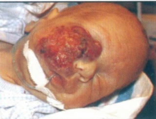
🔍 Key clinical features:
- ✓ Bright red, protuberant mass involving the periocular region
- ✓ Eyelid involvement – obstructing the visual axis
- ✓ Rapid growth – typical of proliferating infantile hemangioma
- ✓ Risk of amblyopia (deprivation or strabismic)
- ✓ High-risk hemangioma – requires urgent treatment
📋 Clinical scenario (examiner prompt)
A 3‑month‑old infant presents with a rapidly growing red mass on the left side of the face, first noticed at 2 weeks of age. The lesion now involves the upper and lower eyelids, obstructing the visual axis. The infant is otherwise healthy and feeding well. The parents are concerned about the “bump” and whether the baby can see.
🧑⚕️ Examiner tasks · TOACS
1. Identify the diagnosis from the clinical image and history.
2. Describe the clinical features (rapid growth, periocular involvement, visual axis obstruction).
3. Explain the pathophysiology (infantile hemangioma, GLUT-1+, proliferative phase).
4. Discuss management (urgent propranolol, ophthalmology, PHACES evaluation).
🎯 Expected answers (for examiners)
- • Diagnosis: Periocular infantile hemangioma (high-risk)
- • Clinical features: Bright red mass, eyelid involvement, visual axis obstruction, rapid growth
- • Pathophysiology: GLUT-1+ endothelial proliferation; proliferative phase in first months
- • Management: Oral propranolol (2‑3 mg/kg/day), ophthalmology (urgent), MRI/MRA if PHACES suspected
⚡ Quick FCPS‑style MCQ
A 3‑month‑old with a periocular hemangioma obstructing the visual axis should be treated with:
A. Observation only B. Oral propranolol C. Topical timolol D. Surgical excision📌 Topic summary ·
GLUT-1+, proliferates first 6‑9 months
Periocular, airway, large facial, PHACES
Oral propranolol (2‑3 mg/kg/day)
Posterior fossa, Arterial, Coarctation, Eye, Sternal
Eyelid obstruction → urgent treatment
Essential for visual outcome
| Feature | Periocular Hemangioma |
|---|---|
| Onset | First weeks, rapid growth in proliferative phase |
| Risk | Amblyopia (deprivation, strabismus), astigmatism |
| Treatment | Oral propranolol (first‑line) |
| Ophthalmology | Urgent referral, monitor for visual axis obstruction |
| PHACES | Evaluate if segmental, >5cm |