🩻 TOACS FCPS Station · Neonatal Skin: Mongolian Spot
📷 Clinical Photograph
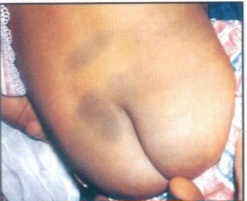
🔍 Key clinical features:
- ✓ Blue-gray to slate-black macular patch
- ✓ Located on the sacrum, buttocks, and lower back
- ✓ Present at birth or appears shortly after
- ✓ No texture change – flat, non-palpable
- ✓ Benign – fades in early childhood
📋 Clinical scenario (examiner prompt)
A 2‑day‑old term infant is noted to have a blue‑gray patch on the sacrum and buttocks during the routine newborn examination. The lesion is flat and non‑palpable. The infant is otherwise healthy, feeding well, and has no other skin lesions. The parents are concerned about the “bruise” on their baby’s back.
🧑⚕️ Examiner tasks · TOACS
1. Identify the diagnosis from the clinical image and history.
2. Describe the clinical features (blue-gray macule, sacral location, flat).
3. Explain the pathophysiology (dermal melanocytes, neural crest migration).
4. Discuss management and parental reassurance (benign, fades with age).
🎯 Expected answers (for examiners)
- • Diagnosis: Mongolian spot (congenital dermal melanocytosis)
- • Clinical features: Blue-gray to slate-black flat macule, sacral/gluteal, present at birth
- • Pathophysiology: Dermal melanocytes trapped in dermis during neural crest migration
- • Management: Reassurance, document in medical record, no treatment needed
⚡ Quick FCPS‑style MCQ
A blue-gray macular patch on the sacrum of a healthy newborn is most likely:
A. Mongolian spot (congenital dermal melanocytosis) B. Bruising from birth trauma C. Nevus simplex (salmon patch) D. Port-wine stain📌 Topic summary ·
Congenital dermal melanocytosis
Present at birth
Blue-gray to slate-black macule
Sacrum, buttocks, lower back
Dermal melanocytes (neural crest)
Fades in early childhood
| Condition | Key Feature | Management |
|---|---|---|
| Mongolian spot | Blue-gray sacral macule, flat | Reassurance, document |
| Bruising | Varies in color, tender, changes over time | Evaluate for trauma/abuse |
| Nevus simplex | Pink, blanching, face/nape | Reassurance |
| Port-wine stain | Pink/red, persistent, unilateral | Laser if needed |